RP-0017
Digital Poster
Filter:
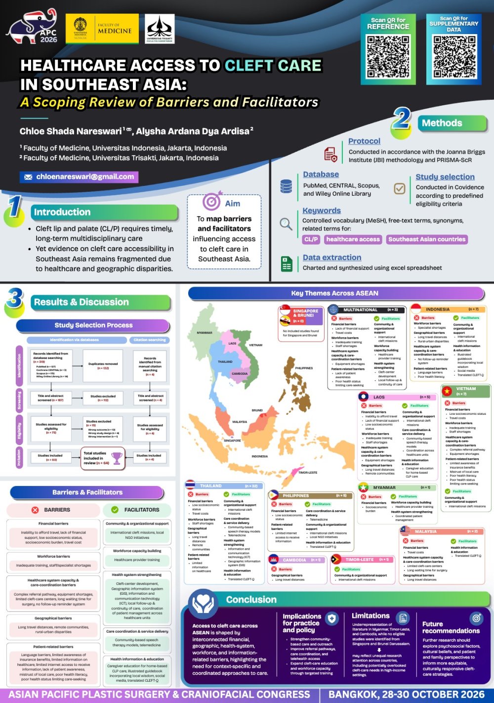
Healthcare Access to Cleft Care in Southeast Asia: A Scoping Review of Barriers and Facilitators RP-0017
Chloe Shada Nareswari · Research
Introduction: Cleft lip and palate (CL/P) requires timely, long-term multidisciplinary care to optimize functional and psychosocial outcomes. Southeast Asia faces substantial disparities in healthcare resources and geographic access, yet evidence on cleft care accessibility across the region remains fragmented. This scoping review mapped barriers and facilitators influencing access to cleft care in Southeast Asia.
Methods: This scoping review was conducted in accordance with the Joanna Briggs Institute (JBI) methodology and PRISMA-ScR. A comprehensive search of PubMed, CENTRAL, Scopus, and Wiley Online Library was performed using controlled vocabulary (MeSH where applicable), free-text terms, synonyms, and related terms for CL/P, healthcare access, and Southeast Asian countries. Study selection was conducted in Covidence according to predefined eligibility criteria. Data were charted and synthesized using thematic analysis.
Results: Sixty-four studies were included. Five categories of barriers were identified: financial (travel costs, low socioeconomic status), geographic (long travel distance), health system (challenging referral procedures, prolonged surgical waiting times, inadequate facilities, absence of a follow-up reminder system), workforce (limited trained medical personnel), and information, communication, and health literacy (limited knowledge of available cleft services, language barriers, low health literacy, limited communication skills). Facilitators were grouped into five domains: health information and patient education (social media, guidebooks, translated CLEFT-Q/assessment tools, specialist and peer advice), care coordination and service delivery innovations (telemedicine, community-based speech therapy, speech camps, treatment scheduling method, integrated care and referral systems), workplace capacity building (training programs), health system strengthening (registries, geographic information systems, information and communication technology, healthcare networks), and community and organizational support (NGOs, volunteer mission teams, healthcare insurance).
Conclusion: Access to cleft care in Southeast Asia remains limited by multiple barriers. Improving outcomes requires context-specific strategies, stronger health systems, and regional collaboration. Future research should address psychosocial factors, cultural beliefs, and patient and family perspectives.
View only · Double-click poster to enlarge · Download & print disabled
Loading poster…
RP-0032
- Title
- An Anatomic Investigation of Human Fetal Upper Eyelid
- Author(s)
- Nobuyuki Mitsukawa
- Affiliation
- Department of Plastic and Reconstructive Surgery, Chiba University Graduate School of Medicine
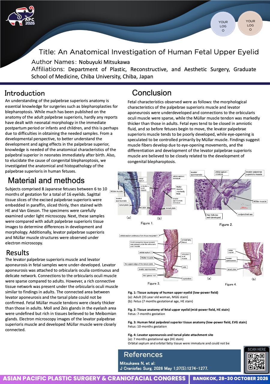
An Anatomic Investigation of Human Fetal Upper Eyelid RP-0032
Nobuyuki Mitsukawa · Research
Introduction: In this study, the authors investigated the anatomic morphology of the upper eyelid in prenatal human fetuses to elucidate the cause of congenital palpebral conditions such as congenital blepharoptosis. Although much has been published on the anatomy of the adult upper eyelid, hardly any reports have dealt with neonatal morphology in the immediate postpartum period or infants and children. From a developmental perspective, to better understand the development and aging effects in the upper eyelid, knowledge is needed of the anatomic characteristics of the upper eyelid in neonates immediately after birth. Also, to elucidate the cause of congenital blepharoptosis, the authors investigated the anatomy of the upper eyelid in human fetuses.
Materials & Methods: Subjects comprised 6 Japanese fetuses between 6 and 10 months of gestation for a total of 12 eyelids. Sagittal tissue slices of the excised upper eyelid were embedded in paraffin, sliced thinly, then stained with HE and Van Gieson. The specimens were carefully examined under light microscopy. Next, these samples were compared with adult upper eyelid tissue images to determine differences in development and morphology.
Results: Fetal characteristics observed were as follows: the morphologic characteristics of the palpebrae superioris muscle and levator aponeurosis were underdeveloped and connections to the orbicularis oculi muscle were sparse, whereas the Müller muscle was markedly thicker than those in adults.
Conclusion: Fetal eyes tend to be closed in amniotic fluid, and so before fetuses begin to move, the levator palpebrae superioris muscle tends to be poorly developed, whereas eye-opening is speculated to be controlled primarily by Müller muscle. Findings suggest muscle fibers develop due to eye-opening movements, and the differentiation and development of the levator palpebrae superioris muscle are believed to be closely related to the development of congenital blepharoptosis.
View only · Double-click poster to enlarge · Download & print disabled
Loading poster…
RP-0033
- Title
- Special distraction osteogenesis before bone grafting for alveolar cleft defects to correct maxillary deformities in patients with bilateral cleft lips and palates
- Author(s)
- Nobuyuki Mitsukawa
- Affiliation
- Department of Plastic, Reconstructive, and Aesthetic Surgery, Graduate School of Medicine, Chiba University
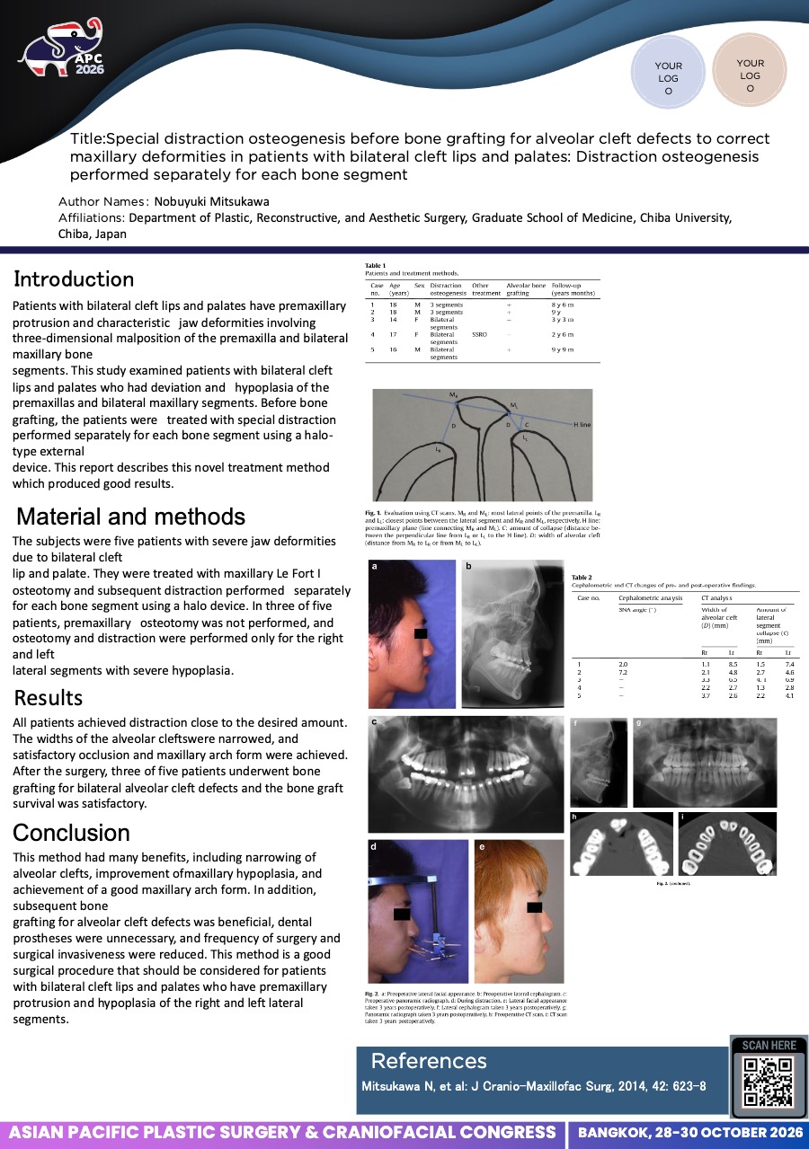
Special distraction osteogenesis before bone grafting for alveolar cleft defects to correct maxillary deformities in patients with bilateral cleft lips and palates RP-0033
Nobuyuki Mitsukawa · Research
Introduction: Patients with bilateral cleft lips and palates have premaxillary protrusion and characteristic jaw deformities involving three-dimensional malposition of the premaxilla and bilateral maxillary bone segments. This study examined patients with bilateral cleft lips and palates who had deviation and hypoplasia of the premaxillas and bilateral maxillary segments. Before bone grafting, the patients were treated with special distraction performed separately for each bone segment using a halo-type external device. This report describes this novel treatment method which produced good results.
Material and methods: The subjects were five patients with severe jaw deformities due to bilateral cleft lip and palate. They were treated with maxillary Le Fort I osteotomy and subsequent distraction performed separately for each bone segment using a halo device. In three of five patients, premaxillary osteotomy was not performed, and osteotomy and distraction were performed only for the right and left lateral segments with severe hypoplasia.
Results: All patients achieved distraction close to the desired amount. The widths of the alveolar clefts were narrowed, and satisfactory occlusion and maxillary arch form were achieved. After the surgery, three of five patients underwent bone grafting for bilateral alveolar cleft defects and the bone graft survival was satisfactory.
Conclusions: This method had many benefits, including narrowing of alveolar clefts, improvement of maxillary hypoplasia, and achievement of a good maxillary arch form. In addition, subsequent bone grafting for alveolar cleft defects was beneficial, dental prostheses were unnecessary, and frequency of surgery and surgical invasiveness were reduced. This method is a good surgical procedure that should be considered for patients with bilateral cleft lips and palates who have premaxillary protrusion and hypoplasia of the right and left lateral segments.
View only · Double-click poster to enlarge · Download & print disabled
Loading poster…
RO-0066
- Title
- Donor-Site-Sparing Biomaterial-Based and Tissue-Engineered Grafts versus Iliac Crest Grafting for Secondary Alveolar Cleft Reconstruction: A Systematic Review and Meta-Analysis of Volumetric Stability and Clinical Outcomes
- Author(s)
- Albertus Andika Wongjaya
- Affiliation
- Department of Plastic Reconstructive and Aesthetic Surgery, Faculty of Medicine, Universitas Lambung Mangkurat, Ulin General Hospital, Banjarmasin, Indonesia
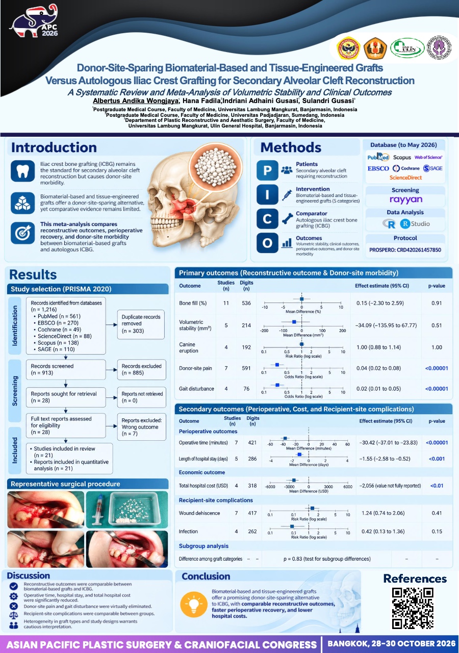
Donor-Site-Sparing Biomaterial-Based and Tissue-Engineered Grafts versus Iliac Crest Grafting for Secondary Alveolar Cleft Reconstruction: A Systematic Review and Meta-Analysis of Volumetric Stability and Clinical Outcomes RO-0066
Albertus Andika Wongjaya · Research
Introduction: Despite decades as the reference standard for secondary alveolar cleft reconstruction, iliac crest bone grafting (ICBG) necessitates a second surgical site with well-documented sequelae. Biomaterial-based and tissue-engineered alternatives have emerged, yet no meta-analysis has synthesized their reconstructive, perioperative, economic, and donor-site morbidity outcomes against ICBG.
Methods: Following PRISMA 2020 (PROSPERO: CRD420261457850), six databases (PubMed, Scopus, EBSCO, ScienceDirect, Cochrane, and Sage) were searched through May 2026. Twenty-one studies (N=774; 348 biomaterial, 426 ICBG) spanning five graft categories were analyzed using DerSimonian–Laird random-effects models; zero-event outcomes were pooled via Peto odds ratio. Risk of bias was assessed with RoB-2 and ROBINS-I; certainty with GRADE framework.
Results: Biomaterial-based and tissue-engineered grafts demonstrated comparable bone fill (k=11; MD +0.15%, 95% CI −2.30 to +2.59; P=0.91; I²=0%), volumetric stability (k=5; MD −34.09 mm³, 95% CI −135.95 to +67.77; P=0.51; I²=0%), and canine eruption (k=4; RR 1.00, 95% CI 0.88–1.14; I²=0%), with no difference among graft categories (P=0.83). Operative time was significantly shorter (k=7; MD −30.42 min, 95% CI −37.01 to −23.83; P<0.00001), as was hospital stay (k=5; MD −1.55 days, 95% CI −2.58 to −0.52; P<0.001). Total hospital cost favored biomaterials despite higher material costs (MD −$2,056; P<0.01). Donor-site pain was virtually absent (0/302 vs 59/289; Peto OR 0.04, 95% CI 0.02–0.08; P<0.00001), as was gait disturbance (0/38 vs 38/38; Peto OR 0.02, 95% CI 0.01–0.05; P<0.00001). Recipient-site complications were equivalent: dehiscence (k=7; RR 1.24, 95% CI 0.74–2.06; P=0.41) and infection (k=4; RR 0.42, 95% CI 0.13–1.36; P=0.15).
Conclusion: Biomaterial-based and tissue-engineered grafts achieve reconstructive parity with autologous ICBG while eliminating donor-site morbidity, shortening perioperative recovery, and reducing hospital expenditure, establishing them as a compelling evidence-based alternative for secondary alveolar cleft reconstruction.
View only · Double-click poster to enlarge · Download & print disabled
Loading poster…
RP-0022
- Title
- TOWARD PRECISION IN MANDIBULAR RECONSTRUCTION: AN UPDATED SYSTEMATIC REVIEW AND META-ANALYSIS OF THREE-DIMENSIONAL VIRTUAL SURGICAL PLANNING VERSUS CONVENTIONAL PLANNING
- Author(s)
- Albertus Andika Wongjaya
- Affiliation
- Departement of Plastic Reconstructive and Aesthatic Surgery, Faculty of Medicine, Universitas Lambung Mangkurat, Ulin General Hospital, Banjarmasin, Indonesia
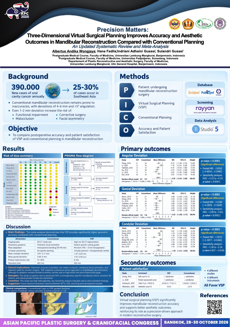
TOWARD PRECISION IN MANDIBULAR RECONSTRUCTION: AN UPDATED SYSTEMATIC REVIEW AND META-ANALYSIS OF THREE-DIMENSIONAL VIRTUAL SURGICAL PLANNING VERSUS CONVENTIONAL PLANNING RP-0022
Albertus Andika Wongjaya · Research
Introduction:
Mandibular reconstruction remains a major challenge in craniofacial surgery because precise
restoration of mandibular anatomy is essential for both functional recovery and facial
symmetry. Conventional planning relies on two-dimensional imaging and intraoperative
freehand techniques, which may limit accurate reproduction of complex three-dimensional
anatomy. However, robust evidence demonstrating whether three-dimensional virtual surgical
planning (VSP) improves reconstruction accuracy and patient-perceived aesthetic outcomes
remains limited. Therefore, this study evaluated the effectiveness of VSP compared with
conventional planning in mandibular reconstruction.
Methods:
A systematic review and meta-analysis were conducted following the PRISMA guidelines.
Searches were performed in PubMed, Scopus, and Cochrane to identify studies comparing
VSP-assisted mandibular reconstruction with conventional surgical planning. Quantitative
outcomes related to postoperative accuracy and facial symmetry, including angular deviation,
gonial positional deviation, and condylar positional deviation, were analyzed in R software
(metafor package). Patient-reported aesthetic outcomes were summarized qualitatively.
Results:
A total of 15 studies were included in the final analysis. The outcomes were grouped into two
categories: quantitative reconstruction accuracy and patient-reported aesthetic outcomes. The
pooled analysis demonstrated significantly lower postoperative angular deviation in the VSP
group (MD −3.95°, 95% CI −5.69 to −2.21; I² = 82.4%). Similarly, VSP significantly reduced
gonial positional deviation (MD −3.22 mm, 95% CI −4.28 to −2.17, I2 = 68.7%) and condylar
positional deviation (MD −1.93 mm, 95% CI −3.31 to −0.55, I2 = 82.8%). Studies evaluating
patient-reported outcomes consistently demonstrated higher aesthetic satisfaction among
patients undergoing VSP-assisted reconstruction.
Conclusion:
Three-dimensional virtual surgical planning significantly improves the accuracy of mandibular
reconstruction and is associated with higher patient-reported aesthetic satisfaction. These
findings support the integration of three-dimensional virtual surgical planning technologies to
achieve more precise, predictable, and patient-centered outcomes in craniofacial reconstructive
surgery.
View only · Double-click poster to enlarge · Download & print disabled
Loading poster…
RP-0025
- Title
- Alveolar Bone Grafting in Patients with Cleft Lip and Palate: An 11 years Retrospective Review from Sarawak General Hospital
- Author(s)
- Fathin alyaa Mohamad farid
- Affiliation
- Sarawak General Hospital, Malaysia
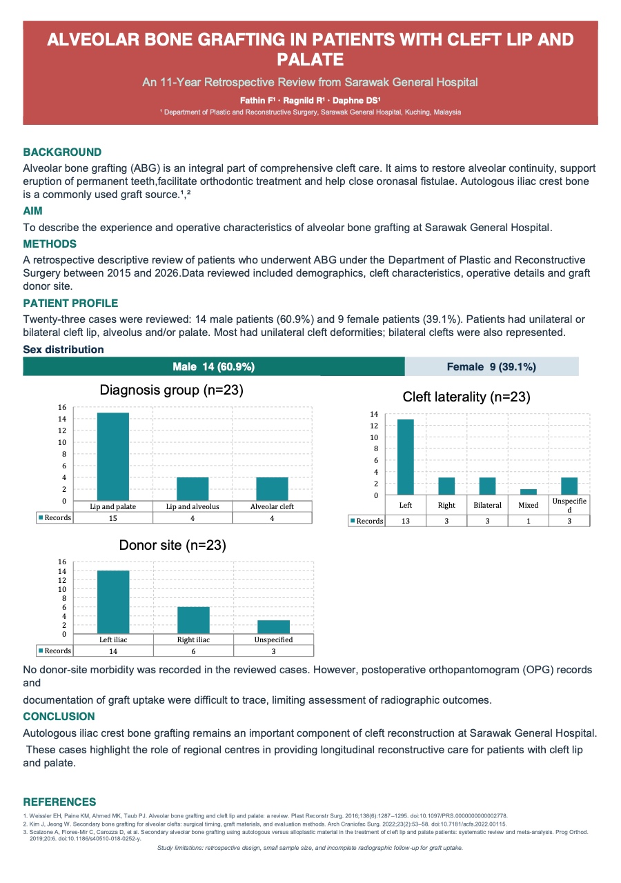
Alveolar Bone Grafting in Patients with Cleft Lip and Palate: An 11 years Retrospective Review from Sarawak General Hospital RP-0025
Fathin alyaa Mohamad farid · Research
Background:
Alveolar bone grafting is an essential component of comprehensive cleft care. It aims to restore alveolar continuity, provide bony support for eruption of the permanent dentition, facilitate orthodontic treatment, and closure of oronasal fistulae. Autologous iliac crest bone remains a commonly used graft source. We describe our experience with alveolar bone grafting at Sarawak General Hospital.
Methods:
A retrospective descriptive review was conducted of patients who underwent alveolar bone grafting under the Plastic and Reconstructive Surgery Department between 2015 and 2026(11years). Data on patient demographics, cleft characteristics, operative details, graft donor site.
Results:
A total of 23 reviewed cases included 9 female patients and 14 male patients with age ranging from 11 years old to 2 years old with unilateral and bilateral cleft lip, alveolus and/or palate. Most patients had unilateral cleft deformities, while bilateral clefts were also represented. Patients underwent alveolar bone grafting using autologous bone harvested predominantly from the anterior iliac crest. Both left and right iliac crest donor sites were utilised according to operative requirements.
Several patients underwent concomitant procedures, including lip revision, correction of nostril, and dermal fat grafting. Operative procedures were performed electively under general anaesthesia. Recorded operative durations varied from approximately 71 to 169 minutes. The cases demonstrated no donor site morbidity. however, our limitation was in tracing the OPG / bone graft uptake.
Conclusion:
Alveolar bone grafting using autologous iliac crest bone remains an important component of cleft reconstruction at our centre. This study reflects the role of regional centres in providing longitudinal reconstructive care for patients with cleft lip and palate.
View only · Double-click poster to enlarge · Download & print disabled
Loading poster…
RP-0015
- Title
- Orthodontic Management Protocols in Orthognathic Surgery for Cleft and Non-Cleft and Their Treatment Outcomes: A Systematic Review
- Author(s)
- WAN SALBIAH WAN ABDUL RAZAK
- Affiliation
- University Technology MARA / Oral Health Division , The Ministry of Health, Malaysia
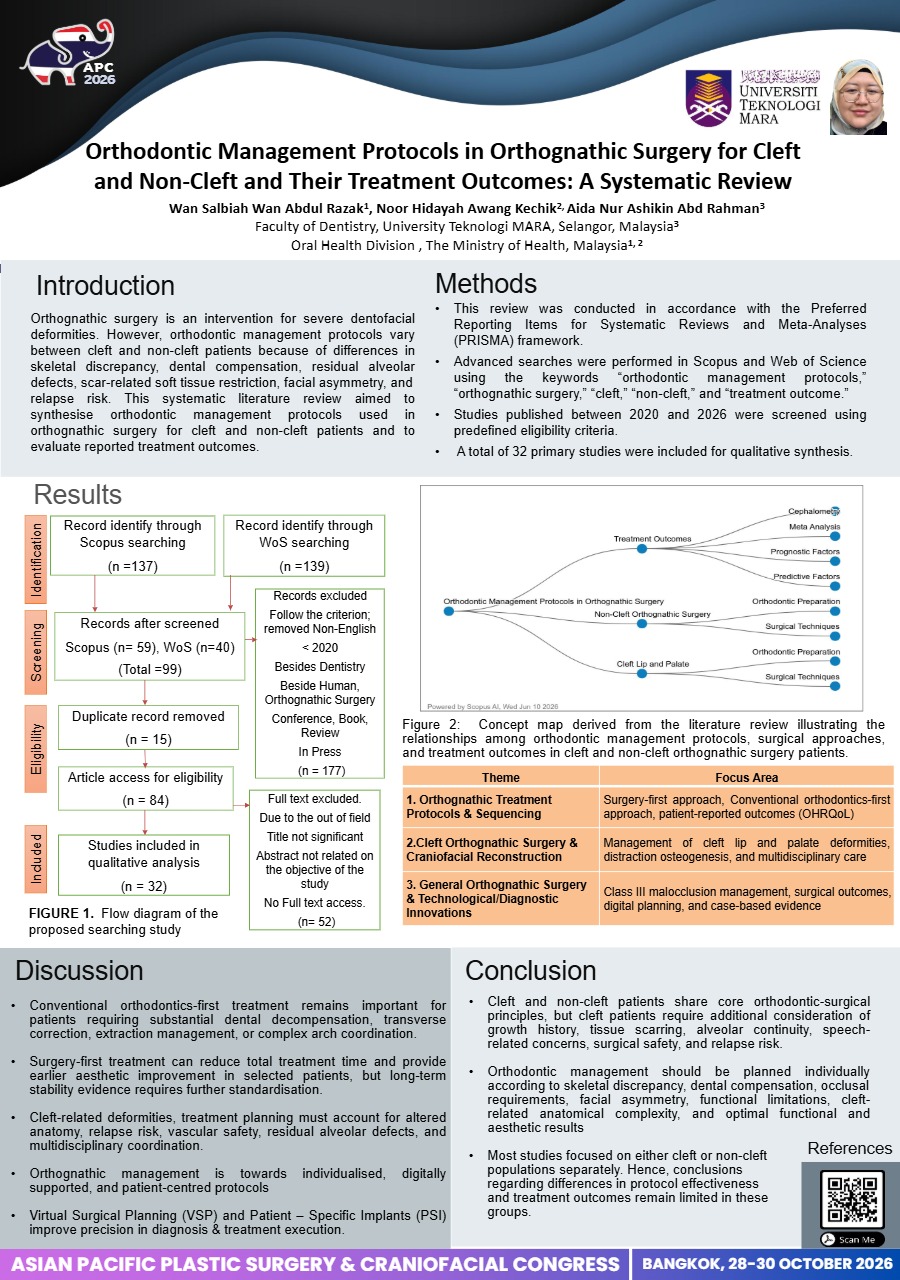
Orthodontic Management Protocols in Orthognathic Surgery for Cleft and Non-Cleft and Their Treatment Outcomes: A Systematic Review RP-0015
WAN SALBIAH WAN ABDUL RAZAK · Research
Background: Orthognathic surgery is an established intervention for severe dentofacial deformities; however, orthodontic management protocols vary between cleft and non-cleft patients because of differences in skeletal discrepancy, dental compensation, residual alveolar defects, scar-related soft tissue restriction, facial asymmetry, and relapse risk. This systematic literature review aimed to synthesise orthodontic management protocols used in orthognathic surgery for cleft and non-cleft patients and to evaluate reported treatment outcomes.
Methods: This review was conducted in accordance with the Preferred Reporting Items for Systematic Reviews and Meta-Analyses (PRISMA) framework. Advanced searches were performed in Scopus and Web of Science using the keywords “orthodontic management protocols,” “orthognathic surgery,” “cleft,” “non-cleft,” and “treatment outcome.” Studies published between 2020 and 2026 were screened using predefined eligibility criteria. A total of 32 primary studies were included for qualitative synthesis.
Results: Three major themes were identified: orthognathic treatment protocols and sequencing; cleft orthognathic surgery and craniofacial reconstruction; and general orthognathic surgery with technological and diagnostic innovations. Conventional orthodontics-first protocols remained important for dental decompensation, arch coordination, extraction management, and correction of complex transverse, vertical, and asymmetrical discrepancies. Surgery-first protocols demonstrated reduced overall treatment duration, earlier facial improvement, and acceptable skeletal stability in selected skeletal Class III cases. In cleft-related deformities, treatment commonly required staged orthodontic preparation, multidisciplinary planning, careful maxillary movement, and management of residual alveolar defects, scar-related tissue limitations, and relapse risk. Digital planning, three-dimensional imaging, computer-aided simulation, three-dimensional printed splints, and clear aligner systems were increasingly applied to improve diagnostic accuracy and surgical precision.
Conclusion: Orthodontic management for orthognathic surgery should be individualised according to skeletal pattern, dental compensation, cleft-related anatomical complexity, functional requirements, and patient-centred outcomes. Further prospective studies with standardised protocols and long-term follow-up are required to strengthen comparative evidence.
View only · Double-click poster to enlarge · Download & print disabled
Loading poster…
RP-0027
- Title
- Three-Dimensional Assessment of Upper Airway Changes After Mandibular Distraction Osteogenesis in Patients With Severe Micrognathia
- Author(s)
- Kahoko Yamada
- Affiliation
- Department of Plastic, Reconstructive, and Aesthetic Surgery, Graduate School of Medicine, Chiba University, Chiba, Japan
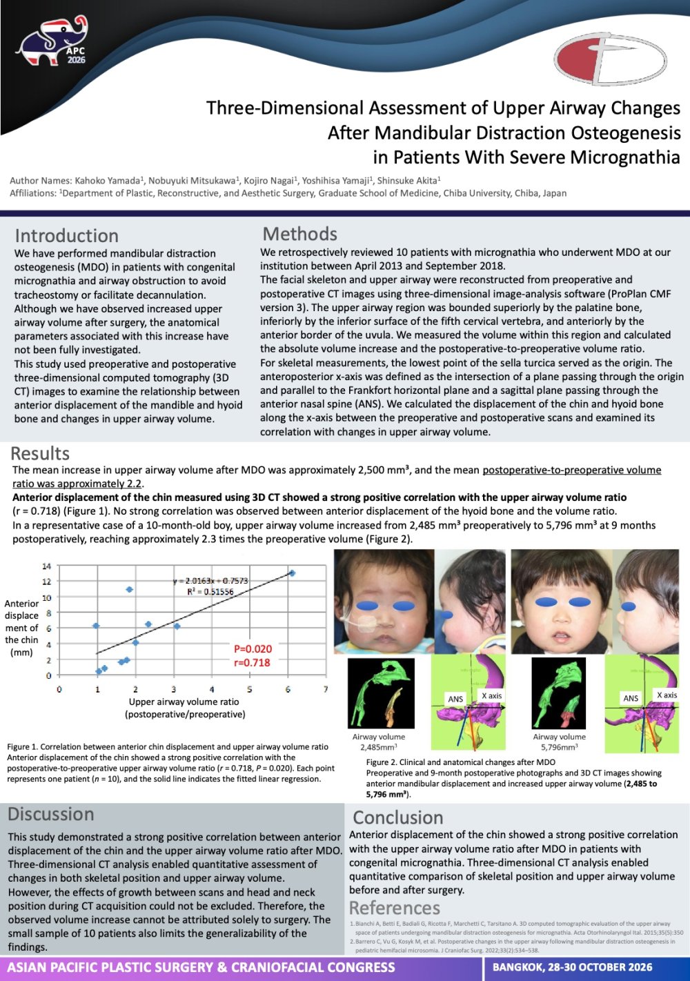
Three-Dimensional Assessment of Upper Airway Changes After Mandibular Distraction Osteogenesis in Patients With Severe Micrognathia RP-0027
Kahoko Yamada · Research
Introduction:
Congenital micrognathia may cause severe upper airway obstruction, and mandibular distraction osteogenesis can facilitate tracheostomy avoidance or decannulation. However, the anatomical changes associated with postoperative airway enlargement remain incompletely understood. This study evaluated upper airway volume before and after mandibular distraction osteogenesis using three-dimensional computed tomography and examined its relationship with anterior displacement of the chin and hyoid bone.
Materials & Methods:
We retrospectively reviewed 10 patients with congenital micrognathia who underwent mandibular distraction osteogenesis at our institution between April 2013 and September 2018. Preoperative and postoperative computed tomography images were reconstructed using three-dimensional image-analysis software. Upper airway volume was calculated within a standardized region defined using the palatal plane, uvular tip, and inferior surface of the fifth cervical vertebra. A three-dimensional coordinate system based on the lowest point of the sella turcica and the Frankfort horizontal plane was used to measure anterior displacement of the chin and hyoid bone. Associations between these displacements and changes in upper airway volume were examined.
Results:
Upper airway volume increased overall after mandibular distraction osteogenesis. The mean absolute increase was approximately 2,500 mm³, and the mean postoperative-to-preoperative volume ratio was approximately 2.2. Anterior displacement of the chin measured by three-dimensional computed tomography showed a strong positive correlation with the proportional increase in upper airway volume (r = 0.718). Hyoid displacement varied considerably among patients and showed no strong correlation with airway volume change.
Conclusion:
Anterior displacement of the chin after mandibular distraction osteogenesis was associated with proportional enlargement of the upper airway. Three-dimensional computed tomography enables quantitative assessment of postoperative airway changes and may complement conventional cephalometric evaluation in patients with severe micrognathia.
View only · Double-click poster to enlarge · Download & print disabled
Loading poster…
RP-0034
- Title
- Evaluation of Alveolar Bone Formation Following Primary Gingivoperiosteoplasty in Patients With Unilateral Complete Cleft Lip and Palate
- Author(s)
- Madoka Sugiyama
- Affiliation
- Department of Plastic and Reconstructive Surgery, Kanagawa Children's Medical Center, Yokohama, Japan
Evaluation of Alveolar Bone Formation Following Primary Gingivoperiosteoplasty in Patients With Unilateral Complete Cleft Lip and Palate RP-0034
Madoka Sugiyama · Research
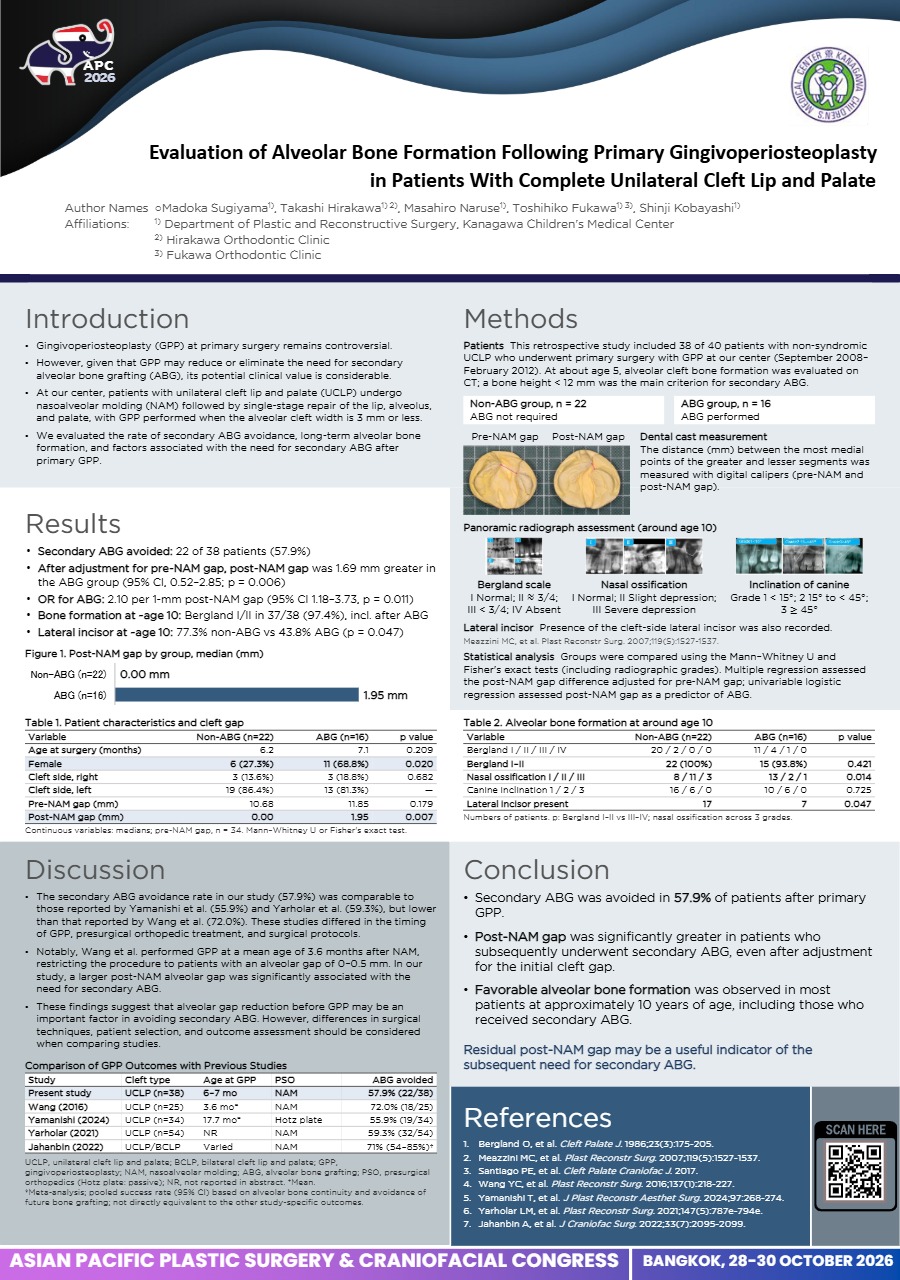
Evaluation of Alveolar Bone Formation Following Primary Gingivoperiosteoplasty in Patients With Unilateral Complete Cleft Lip and Palate
Introduction:
At our center, patients with unilateral complete cleft lip and palate (UCLP) undergo presurgical orthopedics followed by simultaneous primary repair of the lip, alveolus, and palate. Although the role of gingivoperiosteoplasty (GPP) at primary repair remains controversial, successful bone formation may reduce the need for secondary alveolar bone grafting (ABG). This study evaluated alveolar bone formation following primary GPP and the subsequent need for secondary ABG.
Methods:
Forty patients with nonsyndromic UCLP who underwent primary surgery between September 2008 and February 2012 were retrospectively reviewed. The need for secondary ABG was assessed, and alveolar bone formation was evaluated on panoramic radiographs using the Bergland classification.
Results:
Thirty-eight of 40 patients (95.0%) underwent GPP during primary repair. Of these, 22 (57.9%) achieved sufficient alveolar bone formation without secondary ABG, whereas 16 (42.1%) subsequently underwent ABG at a mean age of 6.4 ± 0.7 years. In the non-ABG group, the Bergland classification was type I in 20 patients and type II in 2. In the ABG group, the final classification was type I in 11 patients, type II in 4, and type III in 1. No patient required repeat bone grafting.
Conclusion:
Primary GPP performed with simultaneous lip, alveolar, and palate repair avoided secondary ABG in 57.9% of patients with UCLP. These findings suggest that GPP may reduce the need for secondary bone grafting. Further three-dimensional imaging studies are needed to more accurately characterize alveolar bone formation following GPP
View only · Double-click poster to enlarge · Download & print disabled
Loading poster…
RP-0011
- Title
- Reconstruction with bilateral hatchet flaps for medium sized scalp defects.
- Author(s)
- Junghyun Lee
- Affiliation
- Department of Plastic and Reconstructive Surgery, Hallym University Dongtan Sacred Heart Hospital, Hwaseong, Korea
Reconstruction with bilateral hatchet flaps for medium sized scalp defects. RP-0011
Junghyun Lee · Research
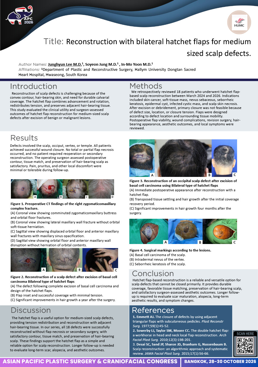
Introduction: Reconstruction of scalp defects is challenging because of the convex contour, hair-bearing skin, and need for durable calvarial coverage. The hatchet flap combines advancement and rotation, redistributes tension, and preserves adjacent hair-bearing tissue. This study evaluated the clinical utility and surgeon-assessed outcomes of hatchet flap reconstruction for medium-sized scalp defects after excision of benign or malignant lesions.
Methods: We retrospectively reviewed 18 patients who underwent hatchet flap-based scalp reconstruction between March 2024 and 2026. Indications included skin cancer, soft-tissue mass, nevus sebaceous, seborrheic keratosis, epidermal cyst, infected cystic mass, and scalp skin necrosis. After excision or debridement, primary closure was not feasible because of defect size, location, or closure tension. Flaps were designed according to defect location and surrounding tissue mobility. Postoperative flap viability, wound complications, revision surgery, hair-bearing appearance, aesthetic outcomes, and local symptoms were reviewed.
Results: Defects involved the scalp, occiput, vertex, or temple. All patients achieved successful wound closure. No total or partial flap necrosis occurred, and no patient required reoperation or secondary reconstruction. The operating surgeon assessed postoperative contour, tissue match, and preservation of hair-bearing scalp as satisfactory. Pain, pruritus, and other local discomfort were minimal or tolerable during follow-up.
Conclusion: Hatchet flap-based reconstruction is a reliable and versatile option for scalp defects that cannot be closed primarily. It provides durable coverage, favorable tissue matching, preservation of hair-bearing scalp, and satisfactory surgeon-assessed aesthetic outcomes. Longer follow-up is required to evaluate scar maturation, alopecia, long-term aesthetic results, and symptom changes.
View only · Double-click poster to enlarge · Download & print disabled
Loading poster…
RO-0056
- Title
- Usefulness of the pedicled latissimus dorsi flap for diverse defective wounds developed in the trunk.
- Author(s)
- Junghyun Lee
- Affiliation
- Department of Plastic and Reconstructive Surgery, Hallym University Dongtan Sacred Heart Hospital, Hwaseong, Korea
Usefulness of the pedicled latissimus dorsi flap for diverse defective wounds developed in the trunk. RO-0056
Junghyun Lee · Research
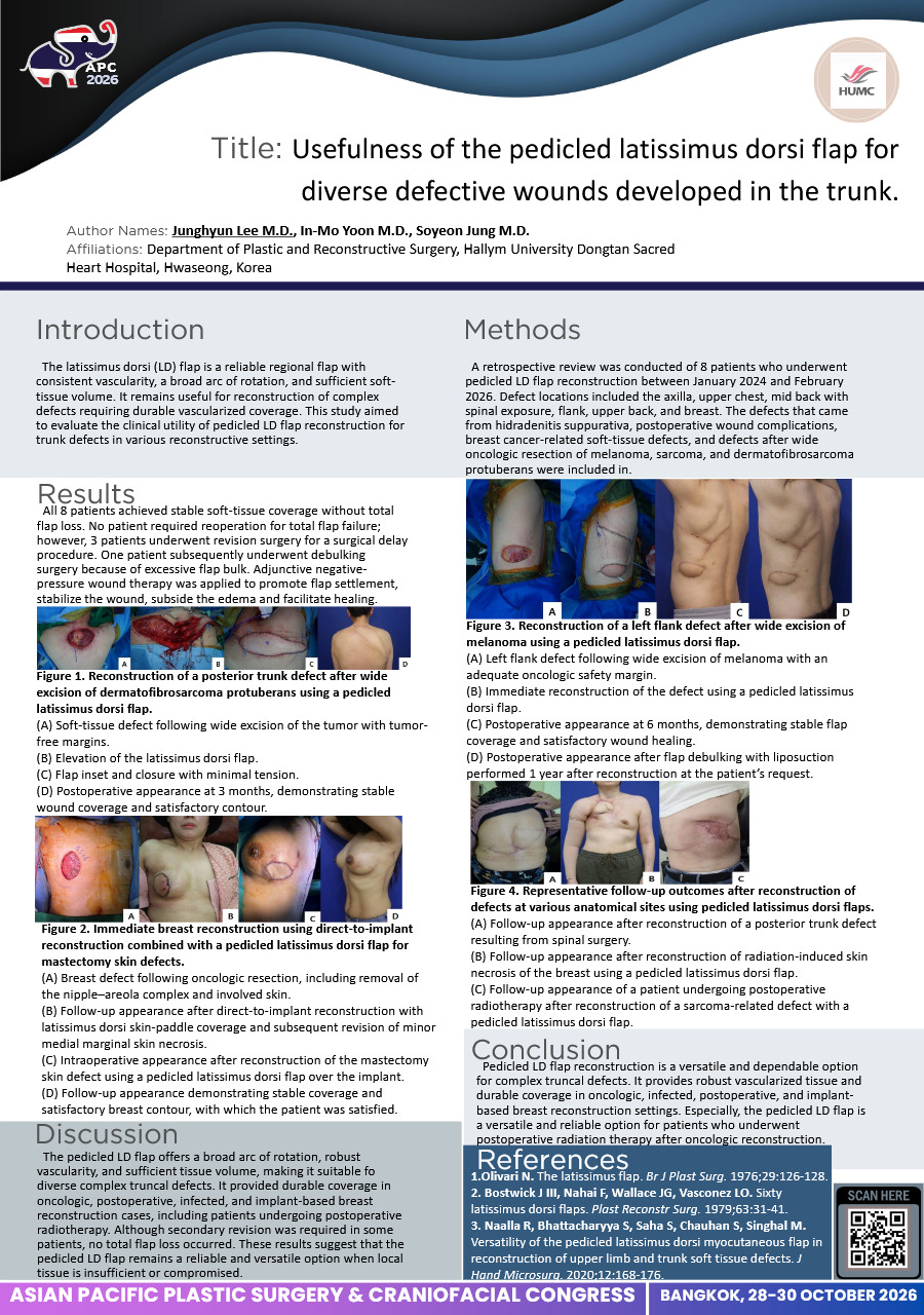
Introduction: The latissimus dorsi (LD) flap is a reliable regional flap with consistent vascularity, a broad arc of rotation, and sufficient soft-tissue volume. It remains useful for reconstruction of complex defects requiring durable vascularized coverage. This study aimed to evaluate the clinical utility of pedicled LD flap reconstruction for trunk defects in various reconstructive settings.
Methods: A retrospective review was conducted of 8 patients who underwent pedicled LD flap reconstruction between January 2024 and February 2026. Defect locations included the axilla, upper chest, mid back with spinal exposure, flank, upper back, and breast. The defects that came from hidradenitis suppurativa, postoperative wound complications, breast cancer-related soft-tissue defects, and defects after wide oncologic resection of melanoma, sarcoma, and dermatofibrosarcoma protuberans were included in.
Results: All 8 patients achieved stable soft-tissue coverage without total flap loss. No patient required reoperation for total flap failure; however, 3 patients underwent revision surgery for a surgical delay procedure. One patient subsequently underwent debulking surgery because of excessive flap bulk. Adjunctive negative-pressure wound therapy was applied to promote flap settlement, stabilize the wound, subside the edema and facilitate healing.
Conclusion: Pedicled LD flap reconstruction is a versatile and dependable option for complex truncal defects. It provides robust vascularized tissue and durable coverage in oncologic, infected, postoperative, and implant-based breast reconstruction settings. Especially, the pedicled LD flap is a versatile and reliable option for patients who underwent postoperative radiation therapy after oncologic reconstruction.
View only · Double-click poster to enlarge · Download & print disabled
Loading poster…
RP-0023
- Title
- A Decade of Cleft Care in Sarawak, Malaysia: A Statewide Registry Review of Cleft Patterns and Surgical Management
- Author(s)
- Daphne Dewi Stephen
- Affiliation
- Hospital Umum Sarawak
A Decade of Cleft Care in Sarawak, Malaysia: A Statewide Registry Review of Cleft Patterns and Surgical Management RP-0023
Daphne Dewi Stephen · Research
Background:
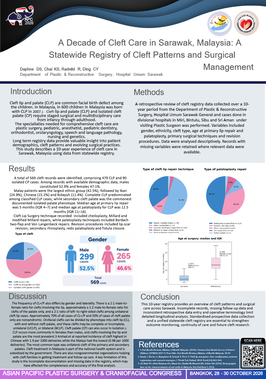
Cleft lip and palate (CLP) and isolated cleft palate (CP) require staged surgical and multidisciplinary care from infancy through adulthood. Long-term registry data provide valuable insight into patient demographics, cleft patterns and evolving surgical practices. This study describes a 10-year experience of cleft care in Sarawak, Malaysia, using data from a statewide cleft registry.
Methods:
A retrospective review of cleft registry data collected over a 10-year period from Sarawak General Hospital/PJS and divisional hospitals in Miri, Bintulu, Sibu and Sri Aman was performed. Variables included gender, ethnicity, cleft type, age at primary lip repair and palatoplasty, primary surgical techniques and revision procedures. Data were analysed descriptively. Records with missing variables were retained where relevant data were available.
Results:
A total of 568 cleft records were identified, comprising 478 CLP and 90 isolated CP cases. Among records with available demographic data, males constituted 52.9% and females 47.1%. Malay patients were the largest ethnic group (42.5%), followed by Iban (24.9%), Chinese (15.2%) and Bidayuh (11.4%). Complete CLP predominated among classified CLP cases, while secondary cleft palate was the commonest documented isolated-palate phenotype. Median age at primary lip repair was 5 months (IQR 4–7) and median age at palatoplasty for CLP was 12.5 months (IQR 11–16). The most common cleft lip procedure technique is Modified Millard Lip repair, while palatoplasty techniques include Bardach two-flap and Von Langenbeck repairs. Revision procedures included lip-scar revision, secondary rhinoplasty, redo palatoplasty and fistula closure.
Conclusion:
This 10-year registry provides an overview of cleft patterns and surgical care across Sarawak. Incomplete records, missing follow-up data and inconsistent retrospective data entry and operative terminology limit detailed longitudinal analysis. Standardised prospective data collection and a unified statewide cleft registry are essential to strengthen outcome monitoring, continuity of care and future cleft research.
View only · Double-click poster to enlarge · Download & print disabled
Loading poster…
Page 1 of 2 (24 poster(s))
Double-click or press Esc to close · Scroll to zoom