CP-0019
Digital Poster
Filter:
Less rigid fixation with non - absorbable sutures in sagittal craniosynostosis in single instituition experiences (Hospital Tunku Azizah, Kuala Lumpur , Malaysia) CP-0019
Hamidah Mohd Zainal · Case Study
Introduction:
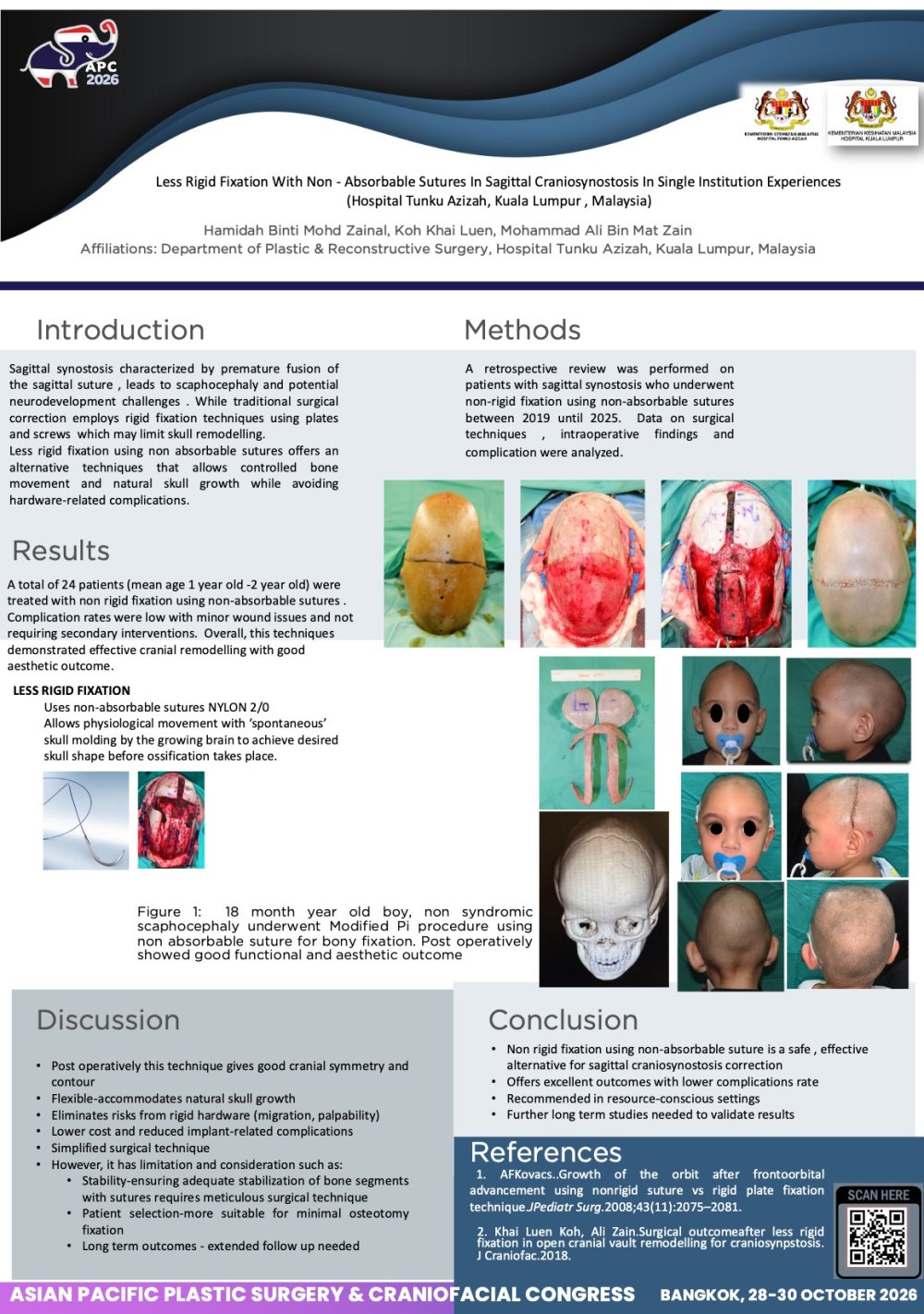
Sagittal Synostosis is the most common craniosynostosis in our center. We performed Cranial vault remodelling using Modified Pi techniques. Due to financial constraints we use non absorbable sutures for bone fixation.
Methods
Retrospective study analysed patient who underwent open anterior cranial vault surgery for saggittal synostosis between . 2017 until 2025 were performed. Demographic data, surgical complication, postoperative asthetic outcomes evaluated. Surgical technique using Modified Pi techniques with bitemporal expansion using non absorbable sutures.
Results:
25 patients of sagittal synostosis with mean age 1.7 month operated using modified pi technique and all cases use nylon 2/0 for bone fixation. Non of our series of case has complication related to suture used, ie: suture extrusion, infection or wound breakdown. All the cases gives good aesthetic outcome.
Conclusion:
Our technique of open cranial vault reconstruction with non absorbabble sutures nylon 2/0 as sole methods of fixation has resulted in good aesthethic outcome with no reoperation and complication rate.
View only · Double-click poster to enlarge · Download & print disabled
Loading poster…
CP-0015
- Title
- LOWER-LID SWITCH FLAP TO MAINTAIN FUNCTION AND AESTHETIC RESULTS OF UPPER EYELID AFTER FULL-THICKNESS WIDE EXCISION OF SQUAMOUS CELL CARCINOMA
- Author(s)
- Faustine Emanuela
- Affiliation
- Plastic Reconstructive and Aesthetic Surgery Unit, Pusat Angkatan Laut dr. Ramelan Hospital, Surabaya, Indonesia
LOWER-LID SWITCH FLAP TO MAINTAIN FUNCTION AND AESTHETIC RESULTS OF UPPER EYELID AFTER FULL-THICKNESS WIDE EXCISION OF SQUAMOUS CELL CARCINOMA CP-0015
Faustine Emanuela · Case Study
Background: Eyelid tumors are relatively rare and heterogeneous. Malignant eyelid tumors tend to display aggressive histopathological behavior, complete surgical excision is essential in their management. Achieving maximal preservation of both cosmetic appearance and function remains a challenging task even for experienced surgeons.
Objective: To describe a two-stage lower-lid switch flap technique for reconstructing the upper eyelid and perserving its function after full-thickness wide excision of squamous cell carcinoma.
Methods: A case report of a 45-year-old man with squamous cell carcinoma of the right upper eyelid.
Result: Reconstruction after full-thickness excision involving more than 75% of the upper eyelid must not only close the defect but also preserve eyelid function. The porcedure was performed in two stages: the lower-eyelid flap was first attached to the upper-eyelid defect, and the flap was the devided after 21 days. Ini ths case, the defect was closed using a lower-lid switch flap based on the marginal arcade, a technique chosen to preserve the tarsal plate and levator palpebrae muscle so that eyelid openinig and closing remained functional. This approach yielded good functional and aesthetic outcomes without complications.
Conclusion: Malignant neoplasms are a leading indication for reconstructive surgery in the periorbital region. Closing an upper-eyelid defect without reconstructing the levator palpebrae muscle is restored and the tarsus is preserved as the lid margin, thereby maintaining eyelid function.
View only · Double-click poster to enlarge · Download & print disabled
Loading poster…
CP-0031
- Title
- Orbital Reconstruction Using Dermal Fat Graft After Removal of Magnetic Orbital Prosthesis
- Author(s)
- Nobuyuki Mitsukawa
- Affiliation
- Department of Plastic, Reconstructive, and Aesthetic Surgery, Graduate School of Medicine, Chiba University
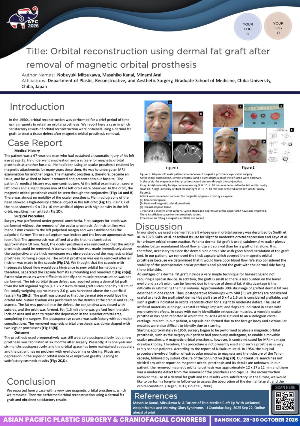
Orbital Reconstruction Using Dermal Fat Graft After Removal of Magnetic Orbital Prosthesis CP-0031
Nobuyuki Mitsukawa · Case Study
Introduction: We present a case of dermal fat graft for eye socket reconstruction after the removal of magnetic orbital prosthesis.
Case Report :A 68-year-old male who had a damage to the left eye by trauma more than 40 years ago, and the eyeball was removed and magnetic orbital prosthesis surgery was performed. The patient was using the magnetic orbital prosthesis for many years, but upon receiving MRI examination, the magnet became an obstacle, and came to our department to remove it. At the first visit, pelvic drops on the affected side and mild depression of the upper eyelid were observed, and the magnetic orbital prosthesis was transparent through the conjunctiva. Under general anesthesia, made incision horizontally to the conjunctiva and removed magnetic orbital prosthesis. After removing the magnetic orbital prosthesis, the dermal fat collected from the groin was transplanted excessively to the tissue defect, and the wound was closed. And also the fat injection was performed to the upper eyelid.
Results/Conclusion: 2 years after the operation, condition of the dermal fat graft is stable, there is no problem wearing the new orbital prosthesis.
View only · Double-click poster to enlarge · Download & print disabled
Loading poster…
CP-0018
- Title
- The Role of Postoperative Nasal Retainer in Achieving Nasal Symmetry after Modified Subunit Cheilonasorraphy: A Case Series
- Author(s)
- Faustine Emanuela
- Affiliation
- Pusat Angkatan Laut dr. Ramelan Hospital, Surabaya, Indonesia
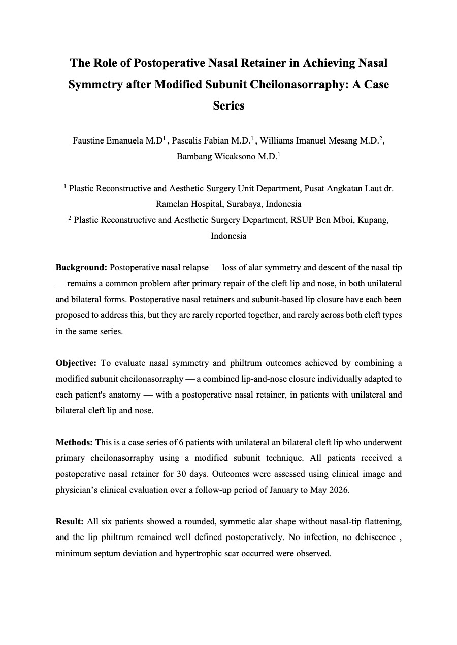
The Role of Postoperative Nasal Retainer in Achieving Nasal Symmetry after Modified Subunit Cheilonasorraphy: A Case Series CP-0018
Faustine Emanuela · Case Study
Background: Postoperative nasal relapse — loss of alar symmetry and descent of the nasal tip — remains a common problem after primary repair of the cleft lip and nose, in both unilateral and bilateral forms. Postoperative nasal retainers and subunit-based lip closure have each been proposed to address this, but they are rarely reported together, and rarely across both cleft types in the same series.
Objective: To evaluate nasal symmetry and philtrum outcomes achieved by combining a modified subunit cheilonasorraphy — a combined lip-and-nose closure individually adapted to each patient's anatomy — with a postoperative nasal retainer, in patients with unilateral and bilateral cleft lip and nose.
Methods: This is a case series of 6 patients with unilateral an bilateral cleft lip who underwent primary cheilonasorraphy using a modified subunit technique. All patients received a postoperative nasal retainer for 30 days. Outcomes were assessed using clinical image and physician’s clinical evaluation over a follow-up period of January to May 2026.
Result: All six patients showed a rounded, symmetic alar shape without nasal-tip flattening, and the lip philtrum remained well defined postoperatively. No infection, no dehiscence , minimum septum deviation and hypertrophic scar occurred were observed.
Conclusion: Combining a modified-subunit cheilonasorraphy technique with postoperative nasal retainer appears promising for perserving nasal shape and philtrum contour in primary repair to unilateral and bilateral cleft lip. Controlled comparative studies with larger samples and objective measurements are needed to confirm the advantage of this technique over conventional approaches.
View only · Double-click poster to enlarge · Download & print disabled
Loading poster…
CP-0017
- Title
- When the Eyelids Heal Toghether: Two-Lamellar Reconstruction of a Rare Post-Traumatic Medial Canthal Ankyloblepharon
- Author(s)
- Faustine Emanuela
- Affiliation
- Pusat Angkatan Laut Dr. Ramelan Hospital, Surabaya, Indonesia
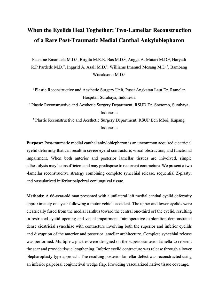
When the Eyelids Heal Toghether: Two-Lamellar Reconstruction of a Rare Post-Traumatic Medial Canthal Ankyloblepharon CP-0017
Faustine Emanuela · Case Study
Purpose: Post-traumatic medial canthal ankyloblepharon is an uncommon acquired cicatricial eyelid deformity that can result in severe eyelid contracture, visual obstruction, and functional impairment. When both anterior and posterior lamellar tissues are inivolved, simple adhesiolysis may be insufficient and may predispose to recurrent contracture. We present a two -lamellar reconstrucitve strategy combining complete synechial release, sequential Z-plasty, and vascularized iniferior palpebral conjungtival tissue.
Methods: A 66-year-old man presented with a unilateral left medial canthal eyelid deformity approximately one year following a motor vehicle accident. The upper and lower eyelids were cicatrically fused from the medial canthus toward the central one-third orf the eyelid, resulting in restricted eyelid opening and visual impairment. Intraoperative exploration demonstrated dense cicatricial synechiae with contracture involving both the superior and inferior eyelids and disruption of the anterior and posterior lamellar architecture. Complete synechial release was performed. Multiple z-plasties were designed on the superior/anterior lamella to reorient the scar and provide tissue lengthening. Inferior eyelid contracture was release through a lower blepharoplasty-type approach. The resulting posterior lamellar defect was reconstructed using an inferior palpebral conjunctival wedge flap. Providing vascularized native tissue coverage.
Result: The procedure restored separation of the upper and lower eyelids re-established the medial canthal architecture. Postoperatively, the patient achieved eyelid opening comparable to the contralateral eye with satisfactory eyelid closure. No wound infection or dehiscence occurred. The patient reported satisfactory functional and aesthetic improvement with a marked improvement in quality of life.
Conclusion: Post-traumatic medial canthal ankyloblepharon should be considered a complex three-dimensional cicatricial deformity rather than a simple eyelid adhesion. Successful reconstruction requires simultaneous correction of the contractile forces and restoration of the ephitelialized surfaces of both eyelid lamellae. The combiniation of synechial release, Z-plasty-based anterior lamellar lengthening, and vascularized conjunctival resurfacing provides a tissue-perservinig release-reorient-resurface stratefy for restoring eyelid separation and function.
View only · Double-click poster to enlarge · Download & print disabled
Loading poster…
CP-0032
- Title
- Combination of Various Maxillary Distraction Osteogenesis for Patients with Severe Complicated Cleft Maxillary Hypoplasia
- Author(s)
- Nobuyuki Mitsukawa
- Affiliation
- Department of Plastic, Reconstructive, and Aesthetic Surgery, Graduate School of Medicine, Chiba University
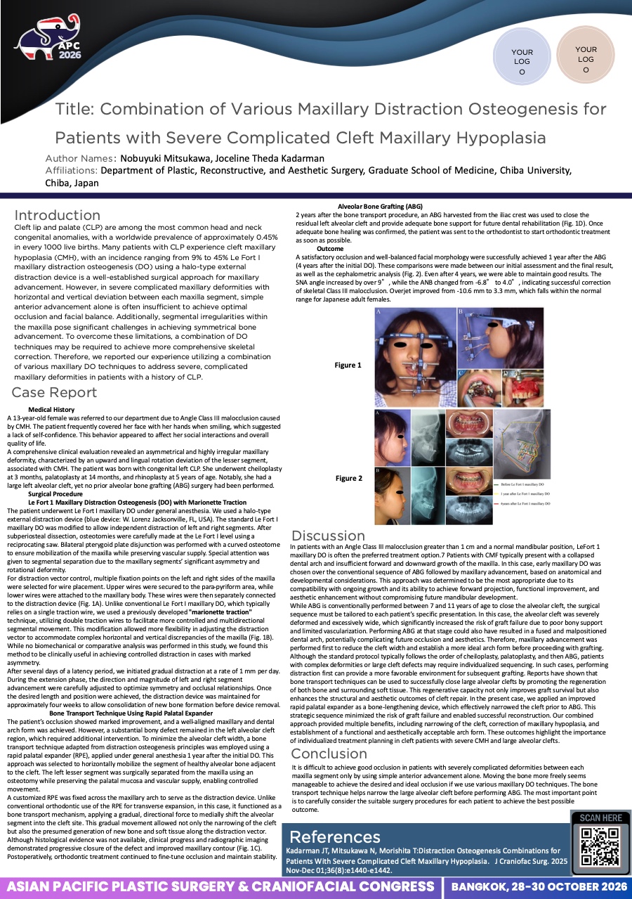
Combination of Various Maxillary Distraction Osteogenesis for Patients with Severe Complicated Cleft Maxillary Hypoplasia CP-0032
Nobuyuki Mitsukawa · Case Study
Cleft maxillary hypoplasia (CMH) is a common sequela in patients with cleft lip and palate (CLP) who have undergone surgical and orthodontic interventions. Le Fort I maxillary distraction osteogenesis (DO) using a halo-type external distraction device is a well-established technique for maxillary advancement. However, simple anterior advancement may not yield satisfactory outcomes in cases involving severe segmental deviations. We reported our success in utilizing a combination of various maxillary DO and bone transport techniques to correct severe, complicated CMH.
Methods: The patient was a 13-year-old girl with Angle Class III malocclusion and a history of congenital left CLP. She had undergone prior corrective surgeries, including cheiloplasty at 3 months of age, palatoplasty at 14 months, and rhinoplasty at 5 years.
Results/Conclusion: Our approach achieved favorable functional and aesthetic outcomes by addressing the complex deformities between maxillary segments. This case highlights the effectiveness of various DO strategies in managing severe, complicated CMH.
View only · Double-click poster to enlarge · Download & print disabled
Loading poster…
CP-0020
- Title
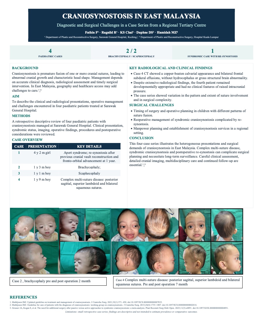
- Craniosynostosis in East Malaysia: Diagnostic and Surgical Challenges in a Case Series from Regional Tertiary Centre
- Author(s)
- Fathin alyaa Mohamad farid
- Affiliation
- Sarawak General Hospital, Malaysia
Craniosynostosis in East Malaysia: Diagnostic and Surgical Challenges in a Case Series from Regional Tertiary Centre CP-0020
Fathin alyaa Mohamad farid · Case Study
Background:
Craniosynostosis is characterised by premature fusion of one or more cranial sutures, resulting in abnormal cranial growth and characteristic skull deformities. Management requires accurate identification of affected sutures, appropriate radiological assessment, and timely surgical intervention. In East Malaysia, geographical and healthcare access considerations may present additional challenges in the diagnosis and management of these uncommon conditions. We describe our experience managing paediatric craniosynostosis at Sarawak General Hospital, highlighting the varied clinical and radiological presentations and surgical challenges encountered.
Methods:
A retrospective descriptive review was conducted of four paediatric patients with craniosynostosis managed at Sarawak General Hospital. Clinical presentation, syndromic status, radiological findings, operative findings, surgical procedures, and postoperative considerations were reviewed.
Four patients were identified, comprising two with brachycephaly and two with scaphocephaly. The cases demonstrated considerable heterogeneity in suture involvement and surgical complexity. A 4-year-2-month-old girl with Apert syndrome developed re-synostosis following previous cranial vault reconstruction and fronto-orbital advancement at one year of age. Reoperation demonstrated fusion of bilateral coronal, sagittal and lambdoid sutures and the anterior fontanelle. Redo cranial vault remodelling using a modified Pi technique with fronto-orbital re-advancement was performed.
A 1-year-3-month-old boy with brachycephaly underwent cranial vault remodelling using a modified Pi technique. A 1-year-1-month-old boy with scaphocephaly and isolated sagittal synostosis underwent cranial vault remodelling. The fourth patient, aged 1 year 9 months, had complex craniosynostosis involving the posterior sagittal, superior lambdoid and bilateral squamous sutures. CT demonstrated a copper-beaten calvarium with bilateral frontal subdural effusions without hydrocephalus. Despite extensive radiological abnormalities, he remained developmentally appropriate without clinical features of raised intracranial pressure.
This case series highlights the challenges of managing craniosynostosis, particularly complex multi-suture disease, syndromic craniosynostosis, and postoperative re-synostosis. Careful clinical assessment, detailed cranial imaging, multidisciplinary care, appropriate surgical timing, and long-term surveillance are essential for optimal management in the regional setting.
View only · Double-click poster to enlarge · Download & print disabled
Loading poster…
CP-0021
- Title
- Proactive Management for Collaborative Cleft Lip and Cleft Palate Care in Upper Northern Part of Thailand
- Author(s)
- Supaksiri Suwiwattana
- Affiliation
- Princess Sirindhorn IT Foundation Craniofacial Center Chiang Mai University
Proactive Management for Collaborative Cleft Lip and Cleft Palate Care in Upper Northern Part of Thailand CP-0021
Supaksiri Suwiwattana · Case Study
Introduction
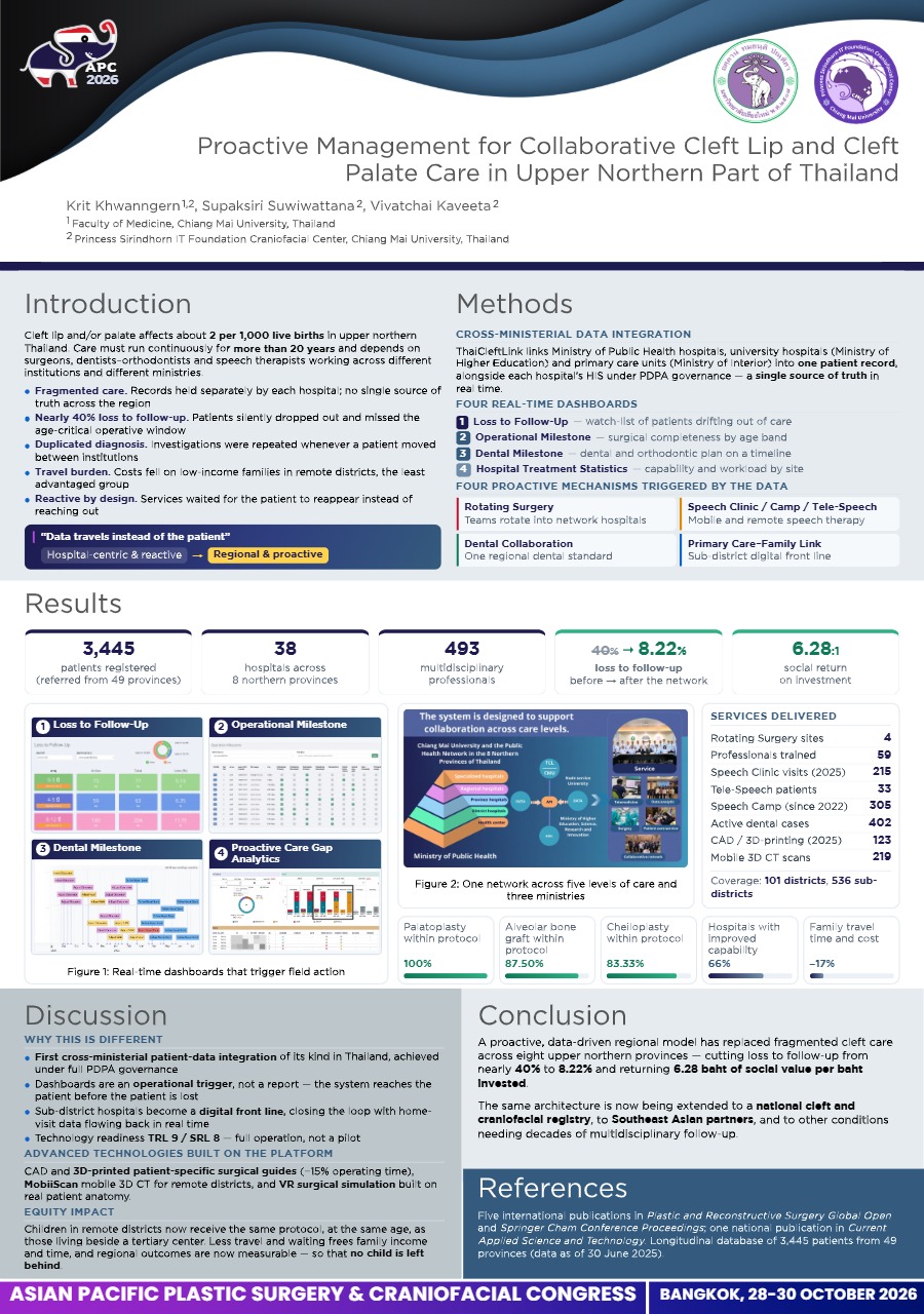
Proactive Management for Collaborative Cleft Lip and Cleft Palate Care in Upper Northern Part of Thailand is a system innovation addressing fragmentation in Thailand’s cleft care. It links university hospitals, Ministry of Public Health hospitals, and community networks into one coordinated, patient-centered system, ensuring continuous care from birth through early adulthood.
Essence of Innovation:
The new data-driven workflow eliminates fragmented care. Built on Chiang Mai University’s multidisciplinary expertise (Medicine, Dentistry, Associated Medical Sciences, Engineer, Information Technology Service Center, and Craniofacial Center), it closes workflow gaps. This collaboration combines network databases with the ThaiCleftLink program to streamline operations, empowering 38 hospitals across 8 northern provinces with standardized care, becomes model for expansion across Southeast Asia. The concept of this innovation system aligns with CMU’s core mission and vision as Thailand’s first regional university, advancing local and national development while supporting its current strategic objectives.
Starting Point:
Began as a CMU multidisciplinary collaboration, then scaled to a regional network linking university, Ministry of Public Health hospitals, and primary care units.
Steps Taken:
Phase 1: Collaborative Network – Established a common vision for coordinated care.
Phase 2: Centralized Data System – Developed the unified ThaiCleftLink platform for patient tracking.
Phase 3: Proactive Management – Conducted a gap analysis, then introduced rotating surgical teams, speech therapy, integrated dental care, telemedicine, and referral systems, dramatically reducing loss-to-follow-up.
Resources Used:
Backed by Chiang Mai University's president, SCFC, and a Ministry of Public Health partnership. Chiang Mai University provided academic/technical expertise, health offices provided personnel, and ITSC provided IT infrastructure.
Impact:
Financial: Patient travel expenses dropped by ~17%. Social/Health Equity: The program supports 2,873 patients via 57 hospitals, and 546 professionals. With the Social Return on Investment (SROI) ratio of 6.28:1, demonstrating high social/economic value.
View only · Double-click poster to enlarge · Download & print disabled
Loading poster…
CP-0023
- Title
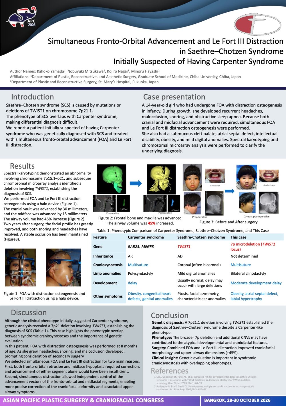
- Simultaneous Fronto-Orbital Advancement and Le Fort III Distraction in Saethre–Chotzen Syndrome Initially Suspected of Having Carpenter Syndrome
- Author(s)
- Kahoko Yamada
- Affiliation
- Department of Plastic, Reconstructive, and Aesthetic Surgery, Graduate School of Medicine, Chiba University, Chiba, Japan
Simultaneous Fronto-Orbital Advancement and Le Fort III Distraction in Saethre–Chotzen Syndrome Initially Suspected of Having Carpenter Syndrome CP-0023
Kahoko Yamada · Case Study
Introduction:
Saethre–Chotzen syndrome is a TWIST1-related craniosynostosis with variable craniofacial and digital manifestations. Phenotypic overlap with other syndromic craniosynostoses may complicate clinical diagnosis. We report a patient initially suspected of having Carpenter syndrome who was genetically diagnosed with Saethre–Chotzen syndrome and treated with simultaneous fronto-orbital advancement and Le Fort III distraction.
Materials & Methods:
We retrospectively reviewed the clinical course of a 14-year-old girl who had undergone fronto-orbital advancement with distraction osteogenesis in infancy. During growth, she developed recurrent headaches, malocclusion, snoring, and obstructive sleep apnea. Because both cranial and midfacial advancement were required, simultaneous fronto-orbital advancement and Le Fort III distraction osteogenesis were performed to allow independent control of the advancement distances and vectors. She also had a submucous cleft palate, atrial septal defect, intellectual disability, obesity, and mild digital anomalies. Spectral karyotyping and chromosomal microarray analysis were performed to clarify the underlying diagnosis.
Results:
The cranial vault and midface were advanced by 30 mm and 15 mm, respectively. Postoperatively, facial profile and occlusion improved, snoring resolved, and recurrent headaches disappeared. These improvements were maintained at 2-year follow-up. Spectral karyotyping demonstrated an abnormality involving chromosome 7p15.1–p21, and subsequent chromosomal microarray analysis identified a deletion involving TWIST1, establishing the diagnosis of Saethre–Chotzen syndrome.
Conclusion:
Simultaneous fronto-orbital advancement and Le Fort III distraction allowed separate control of cranial and midfacial advancement and achieved stable functional and aesthetic improvement. Genetic testing subsequently clarified the diagnosis as Saethre–Chotzen syndrome in this patient with an atypical craniosynostosis phenotype.
View only · Double-click poster to enlarge · Download & print disabled
Loading poster…
CP-0006
- Title
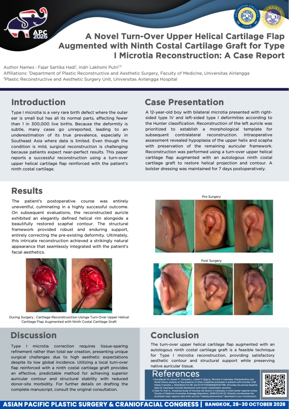
- A Novel Turn-Over Upper Helical Cartilage Flap Augmented with Ninth Costal Cartilage Graft for Type I Microtia Reconstruction: A Case Report
- Author(s)
- Fajar Sartika Hadi
- Affiliation
- Department of Plastic Reconstructive and Aesthetic Surgery, Faculty of Medicine, Universitas Airlangga; Plastic Reconstructive and Aesthetic Surgery Unit, Universitas Airlangga Hospital
A Novel Turn-Over Upper Helical Cartilage Flap Augmented with Ninth Costal Cartilage Graft for Type I Microtia Reconstruction: A Case Report CP-0006
Fajar Sartika Hadi · Case Study
Introduction: Type I microtia is an exceedingly rare congenital auricular anomaly characterized by a hypoplastic auricle with preservation of all anatomical components, with an estimated incidence of fewer than 1 in 300,000 live births. Epidemiological data from Southeast Asia remain limited, restricting accurate assessment of its prevalence in this region. The low reported incidence may reflect the subtle nature of the deformity, which is often unrecognized or not considered severe enough to warrant medical attention, resulting in an iceberg phenomenon where the true disease burden is likely underestimated. Despite its mild external appearance, surgical correction presents unique challenges, as patients seeking reconstruction often have high expectations for achieving a near-normal auricular appearance. We report a case of Type I microtia reconstructed using a turn-over upper helical cartilage flap augmented with an autologous ninth costal cartilage graft.
Case Presentation: A 12-year-old boy with bilateral microtia presented with right-sided type IV and left-sided type I deformities according to the Hunter classification. Reconstruction of the left auricle was prioritized to establish a morphological template for subsequent contralateral reconstruction. Intraoperative assessment revealed hypoplasia of the upper helix and scapha with preservation of the remaining auricular framework. Reconstruction was performed using a turn-over upper helical cartilage flap augmented with an autologous ninth costal cartilage graft to restore helical projection and contour. A bolster dressing was maintained for 7 days postoperatively.
Results : The postoperative course was uneventful, the reconstructed auricle demonstrated a well-defined helical rim, improved scaphal contour, satisfactory structural support, and resulting in a more natural auricular appearance compared with the preoperative deformity.
Conclusion : The turn-over upper helical cartilage flap augmented with an autologous ninth costal cartilage graft is a feasible technique for Type I microtia reconstruction, providing satisfactory aesthetic contour and structural support while preserving native auricular tissue.
View only · Double-click poster to enlarge · Download & print disabled
Loading poster…
CP-0033
- Title
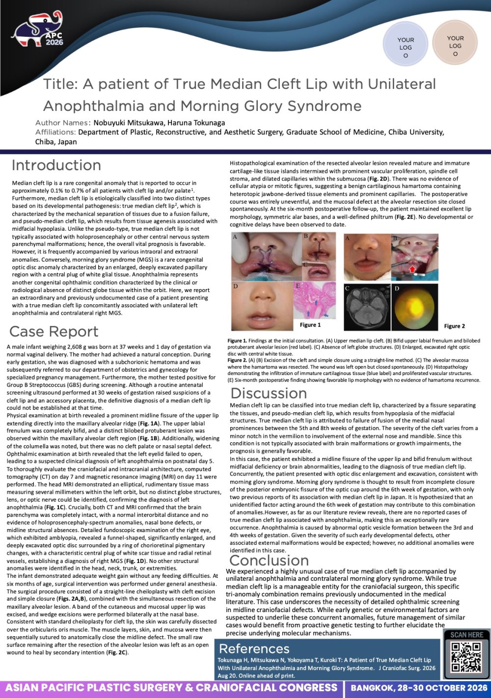
- A Patient of True Median Cleft Lip with Unilateral Anophthalmia and Morning Glory Syndrome
- Author(s)
- Nobuyuki Mitsukawa
- Affiliation
- Department of Plastic, Reconstructive, and Aesthetic Surgery, Chiba University Graduate School of Medicine
A Patient of True Median Cleft Lip with Unilateral Anophthalmia and Morning Glory Syndrome CP-0033
Nobuyuki Mitsukawa · Case Study
Objective:
Median cleft lip is an extremely rare condition, accounting for approximately 0.1–0.7% of cleft lip and palate cases. We report a case of true median cleft lip associated with unilateral anophthalmia and morning glory syndrome.
Case Report:
A male infant weighing 2608 g was born at 37 weeks and 1 day of gestation via normal delivery from a GBS-positive mother. At birth, a fissure was observed in the midline of the upper lip and alveolar ridge, along with a bilobed protuberant lesion in the alveolar cleft region. No brain malformations were detected, and a bifid frenulum was present, confirming the diagnosis of true median cleft lip. Additionally, the patient had right-sided morning glory syndrome and left-sided anophthalmia.
Results:
At six months of age, the patient underwent primary closure of the cleft, wedge excision at the nasal base, and resection of the protuberant lesion in the alveolar cleft region. Histopathological examination revealed that the lesion primarily consisted of immature cartilage-like tissue with vascular proliferation, suggesting a hamartoma caused by heterotopic jawbone-derived tissue. The postoperative course was uneventful, and at two years post-surgery, the patient maintains a favorable lip morphology.
Discussion:
Median cleft lip can be classified into true median cleft lip, characterized by a fissure separating the tissues, and pseudo-median cleft lip, which results from hypoplasia of the midfacial structures. True median cleft lip is attributed to failure of fusion of the medial nasal prominences between the 5th and 8th weeks of gestation. The severity of the cleft varies from a minor notch in the vermilion to involvement of the external nose and mandible. Since this condition is not typically associated with brain malformations or growth impairments, the prognosis is generally favorable.
View only · Double-click poster to enlarge · Download & print disabled
Loading poster…
CP-0030
- Title
- First Successful Le Fort III Distraction in a Patient with Beare-Stevenson Syndrome
- Author(s)
- Nobuyuki Mitsukawa
- Affiliation
- 1) Department of Plastic, Reconstructive, and Aesthetic Surgery, Graduate School of Medicine, Chiba University
First Successful Le Fort III Distraction in a Patient with Beare-Stevenson Syndrome CP-0030
Nobuyuki Mitsukawa · Case Study
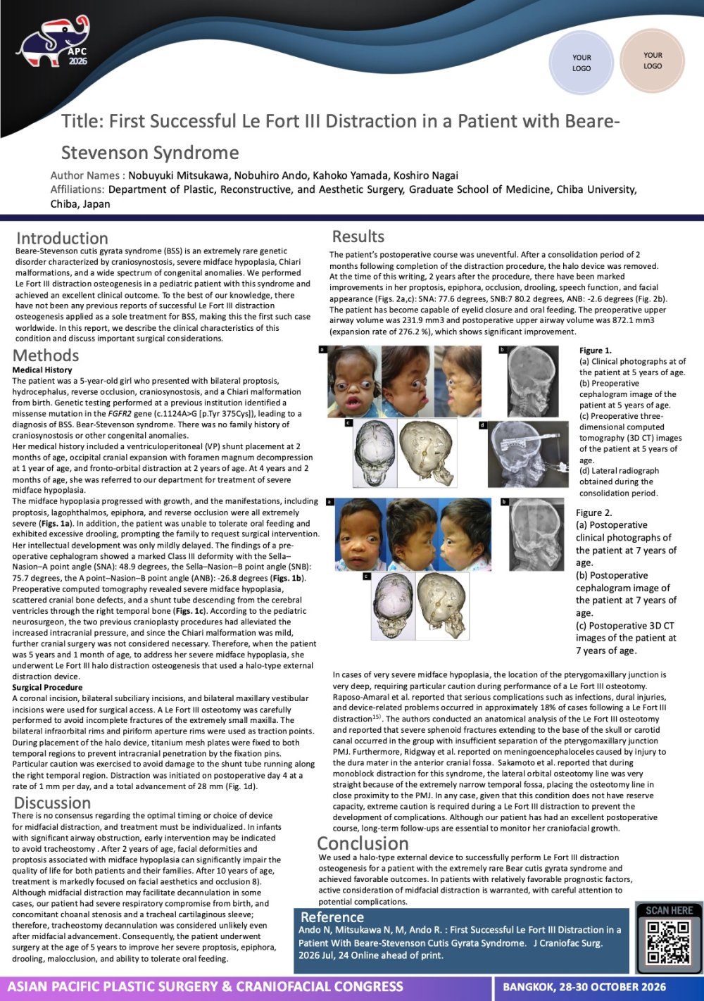
Objective: Beare-Stevenson syndrome is a genetic disorder, caused by FGFR2 gene mutations, characterized by craniosynostosis, midface hypoplasia, Chiari malformation, and other extensive congenital abnormalities. We performed Le Fort III distraction osteogenesis on a girl with this condition and achieved favorable outcomes. This report describes the world's first successful Le Fort III distraction for a patient with Bear-Stevenson syndrome.
Methods: The patient is a 5-year-old girl. She was diagnosed with Beare-Stevenson syndrome shortly after birth and underwent a tracheostomy due to respiratory distress. At 2 month of age, a VP shunt placement was performed, followed by occipital cranial expansion at 1 year and fronto-orbital distraction at 2 years. She was referred to our department for treatment of midface hypoplasia. Clinical findings and imaging studies revealed severe proptosis, very severe midface hypoplasia, and reverse occlusion. We planned Le Fort III distraction using a halo-type external distraction device. Surgery was performed with utmost caution regarding the shunt tube and skull defects.
Results: Postoperatively, an approximately 25 mm distraction was achieved without major complications, 2 years after the operation, significant improvements in proptosis, occlusion, and facial profile were observed, with an excellent clinical course.
Discussion: Beare-Stevenson syndrome is an extremely rare condition presenting with severe midface hypoplasia, with only around 30 reported cases. Due to the poor prognosis associated with respiratory complications, this is, to our knowledge, this case represents the first successful report of Le Fort III distraction. Given the high incidence of Chiari malformations along with the characteristic craniofacial abnormalities, early intervention by both pediatric neurosurgeons and plastic surgeons is crucial. This syndrome presents with severe midface hypoplasia and is associated with poor prognosis due to respiratory complications. However, for cases considered to have a relatively favorable prognosis, Le Fort III distraction should be actively pursued.
View only · Double-click poster to enlarge · Download & print disabled
Loading poster…
Page 1 of 2 (21 poster(s))
Double-click or press Esc to close · Scroll to zoom