CP-0028
Digital Poster
Filter:
A Patient With Mild Trigonocephaly and Concomitant Horizontal Constricting Ring of the Skull CP-0028
Nobuyuki Mitsukawa · Case Study
Introduction:
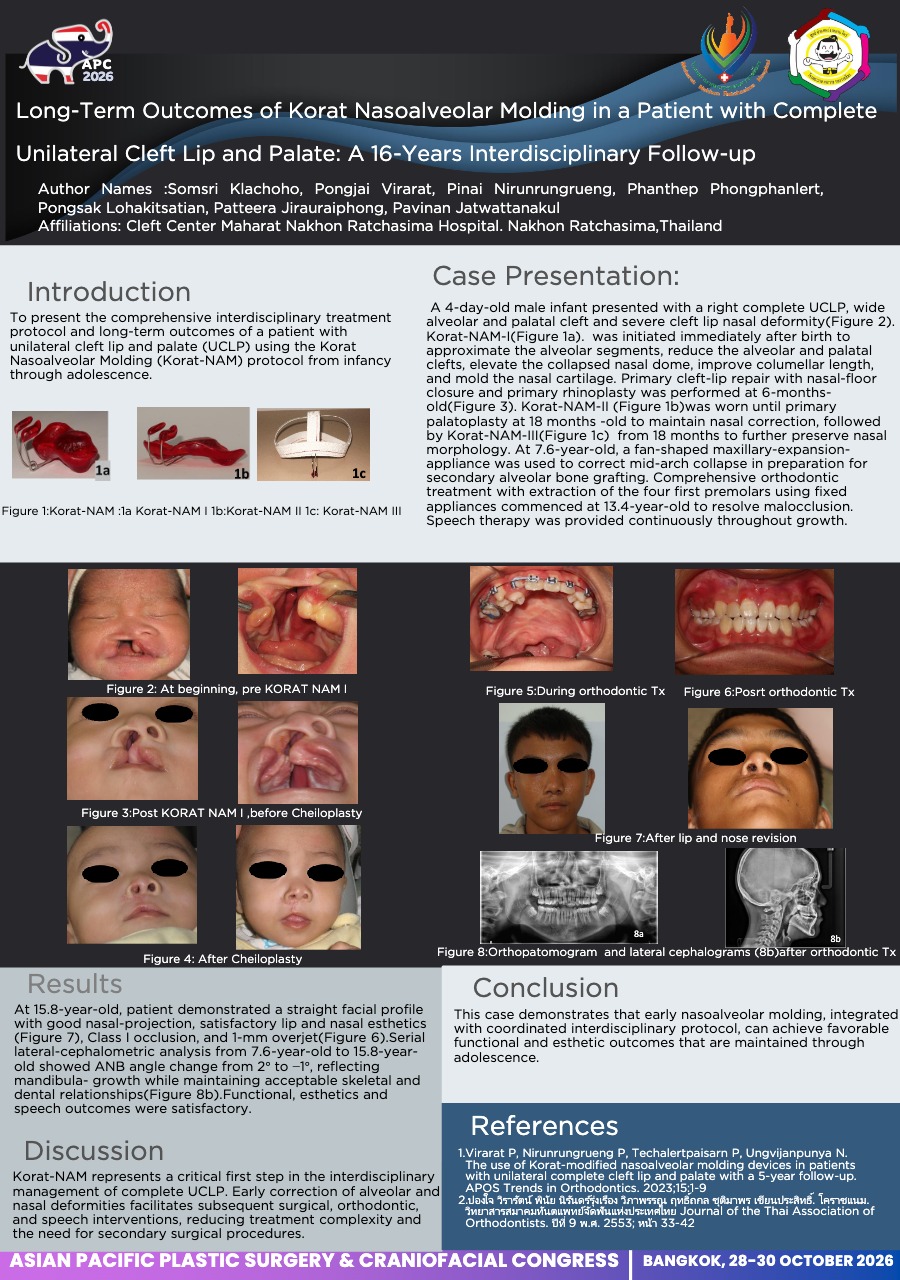
Metopic craniosynostosis causes characteristic cranial deformities such as a keel-shaped forehead, known as trigonocephaly. Mild trigonocephaly refers to cases of metopic craniosynostosis in which the cranial deformity is generally mild. We report a case of mild trigonocephaly accompanied by an atypical horizontal constricting ring of the skull that was successfully treated surgically.
Case Report:
The patient was a 3-month-old boy. CT revealed premature fusion of the metopic suture, and he was referred to our department for surgical treatment. A midline frontal ridge was observed but the keel-shaped forehead was not prominent, and the patient was diagnosed with mild trigonocephaly. Mild depression was observed in the temporal regions, and CT demonstrated a horizontal constricting ring of the skull (circumferential depression) extending approximately three-quarters of the way from the frontal region to both temporal areas. At 11 months of age, one-stage cranial reconstruction was performed for mild trigonocephaly, along with correction of the constricting deformity. Through a coronal incision, the supraorbital bar and frontal bone flap were removed and reshaped. The parietal and temporal bones posterior to the frontal bone flap were barrel-staved, and the depressed bone fragments were elevated to reconstruct the constricted region. Postoperatively, the frontal contour improved, and the constricting ring observed before surgery resolved, resulting in an overall satisfactory cranial shape. One year after surgery, no signs of developmental delay or neurological impairment were observed.
Conclusion:
Cases of nonsyndromic craniosynostosis with a horizontal constricting ring are extremely rare, and to our knowledge, none have been previously reported. Possible etiologies include an amniotic constriction band, a constricting ring caused by squamosal suture fusion as seen in cloverleaf skull, or compensatory deformation associated with restricted cranial growth; however, the exact mechanism remains unclear.
View only · Double-click poster to enlarge · Download & print disabled
Loading poster…
CP-0008
- Title
- Epiphora After Orbital Wall Reconstruction Caused by Migrated Mesh Implant
- Author(s)
- Junghyun Lee
- Affiliation
- Department of Plastic and Reconstructive Surgery, Hallym University Dongtan Sacred Heart Hospital, Hwaseong, Korea
Epiphora After Orbital Wall Reconstruction Caused by Migrated Mesh Implant CP-0008
Junghyun Lee · Case Study
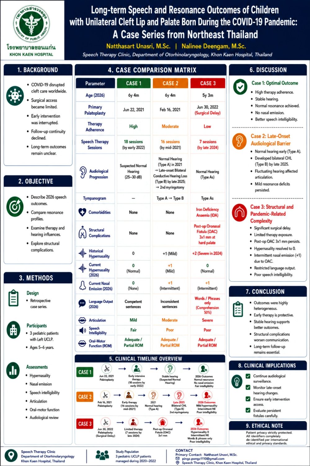
Introduction: Epiphora is an uncommon but clinically significant complication after orbital wall reconstruction. It may occur due to lacrimal drainage disturbance, soft tissue adhesion, or implant-related obstruction. We report a case of postoperative epiphora caused by mesh migration after reconstruction of medial orbital wall and orbital floor fractures.
Methods: A patient sustained left medial orbital wall, orbital floor, and nasal bone fractures following a head-on collision between a bicycle and a vehicle. Extraocular movements were intact, with no diplopia or visual impairment. Orbital wall reconstruction was performed through a transconjunctival-caruncular approach using a bioresorbable mesh which is radiopacity allowing the implant to be clearly visualized on postoperative computed tomography. One month after surgery, the patient developed persistent epiphora. Although ophthalmologic evaluation revealed no definite abnormalities, computed tomography demonstrated displacement of the radiopaque mesh, enabling precise localization of the migrated implant. Based on the correlation between the imaging findings and the patient’s symptoms, surgical exploration and implant complete removal were performed.
Results: The mesh was accurately localized and found to be displaced anteriorly with compressing the lacrimal drainage pathway. Adhesiolysis was performed, and the implant was completely removed. The intraoperative location of the migrated mesh corresponded closely with its position on preoperative computed tomography. At 3 months, the patient reported complete resolution of symptoms without diplopia, visual impairment, or limitation of extraocular movement.
Conclusion: Mesh migration after orbital wall reconstruction may cause delayed epiphora even when ocular motility and vision are normal. Careful evaluation of implant position should be considered in patients with persistent postoperative epiphora. Transcaruncular exploration with implant removal and adhesiolysis may be an effective treatment option.
View only · Double-click poster to enlarge · Download & print disabled
Loading poster…
CP-0009
- Title
- Postoperative Retrobulbar Hematoma With Irreversible Visual Loss After Orbital Fracture Reconstruction In An Older Patient
- Author(s)
- Junghyun Lee
- Affiliation
- Department of Plastic and Reconstructive Surgery, Hallym University Dongtan Sacred Heart Hospital, Hwaseong, Korea
Postoperative Retrobulbar Hematoma With Irreversible Visual Loss After Orbital Fracture Reconstruction In An Older Patient CP-0009
Junghyun Lee · Case Study
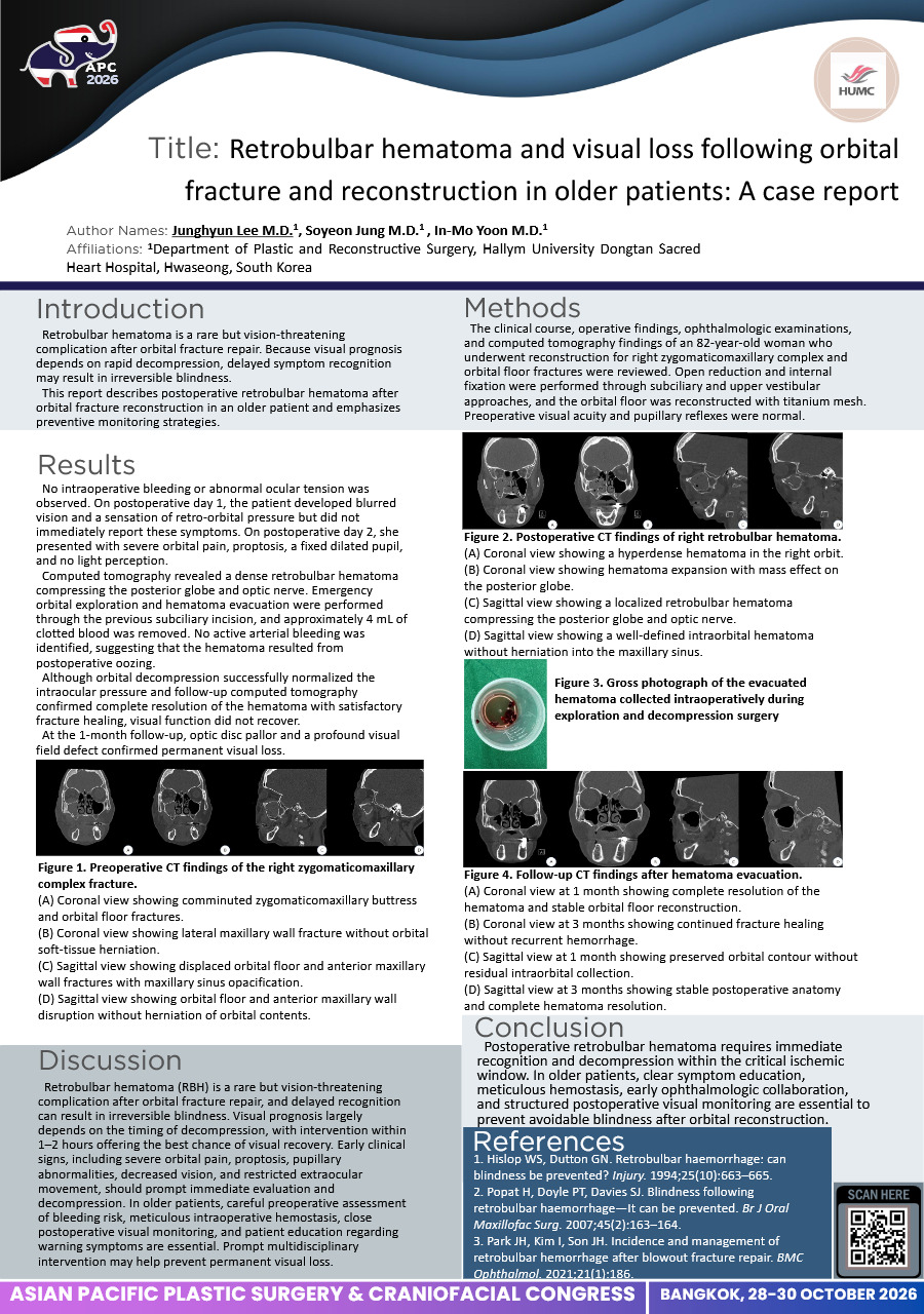
Introduction: Retrobulbar hematoma is a rare but vision-threatening complication after orbital fracture repair. Because visual prognosis depends on rapid decompression, delayed symptom recognition may result in irreversible blindness. This report describes postoperative retrobulbar hematoma after orbital fracture reconstruction in an older patient and emphasizes preventive monitoring strategies.
Methods: The clinical course, operative findings, ophthalmologic examinations, and computed tomography findings of an 82-year-old woman who underwent reconstruction for right zygomaticomaxillary complex and orbital floor fractures were reviewed. Open reduction and internal fixation were performed through subciliary and upper vestibular approaches, and the orbital floor was reconstructed with titanium mesh. Preoperative visual acuity and pupillary reflexes were normal.
Results: No intraoperative bleeding or abnormal ocular tension was observed. On postoperative day 1, the patient developed blurred vision and a sensation of retro-orbital pressure but did not immediately report these symptoms. On postoperative day 2, she presented with severe orbital pain, proptosis, a fixed dilated pupil, and no light perception. Computed tomography revealed a dense retrobulbar hematoma compressing the posterior globe and optic nerve. Emergency orbital exploration and hematoma evacuation were performed through the previous subciliary incision, and approximately 4 mL of clotted blood was removed. No active arterial bleeding was identified, suggesting that the hematoma resulted from postoperative oozing. Although orbital decompression successfully normalized the intraocular pressure and follow-up computed tomography confirmed complete resolution of the hematoma with satisfactory fracture healing, visual function did not recover. At the 1-month follow-up, optic disc pallor and a profound visual field defect confirmed permanent visual loss.
Conclusion: Postoperative retrobulbar hematoma requires immediate recognition and decompression within the critical ischemic window. In older patients, clear symptom education, meticulous hemostasis, early ophthalmologic collaboration, and structured postoperative visual monitoring are essential to prevent avoidable blindness after orbital reconstruction.
View only · Double-click poster to enlarge · Download & print disabled
Loading poster…
CP-0005
- Title
- Modification of Nasoalveolar Molding in a Bilateral Cleft Lip and Palate Patient with a Rotated Premaxilla: A Case Report
- Author(s)
- Thitirath Joongjai
- Affiliation
- Division of Orthodontics, Faculty of Dentistry, Khon Kaen University, Khon Kaen, Thailand
Modification of Nasoalveolar Molding in a Bilateral Cleft Lip and Palate Patient with a Rotated Premaxilla: A Case Report CP-0005
Thitirath Joongjai · Case Study
Introduction: Bilateral cleft lip and palate (BCLP) often involves a severely protruded premaxilla lacking bony continuity with the lateral palatal segments. Conventional nasoalveolar molding (NAM) primarily exerts a single linear retraction vector, which fails to correct premaxillary rotation.
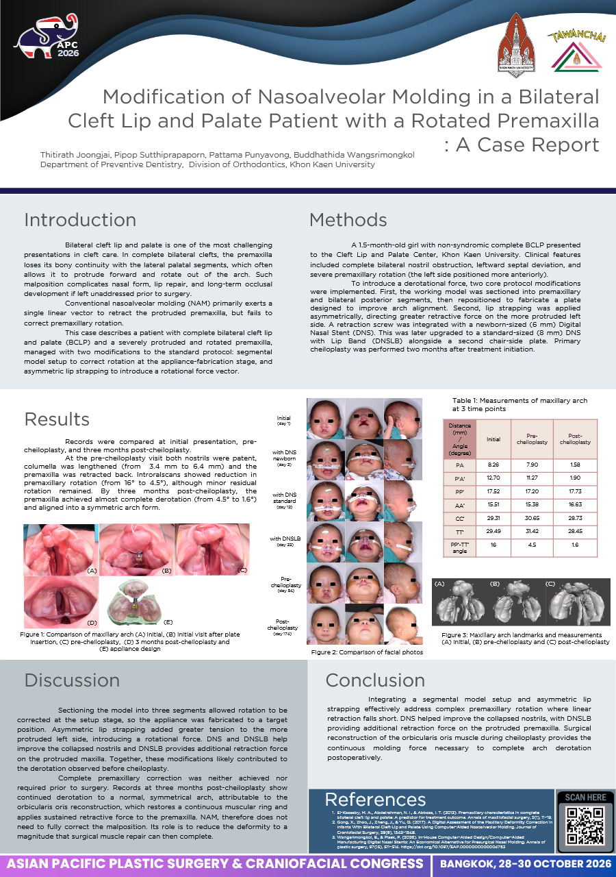
Methods: A 1.5-month-old girl with non-syndromic complete BCLP presented to the Cleft Lip and Palate Center, Khon Kaen University. Clinical features included complete bilateral nostril obstruction, leftward septal deviation, and severe premaxillary rotation (left side positioned more anteriorly). To introduce a derotational force, two core protocol modifications were implemented. First, the working model was sectioned into premaxillary and bilateral posterior segments, then repositioned to fabricate a plate targeted to an improved arch alignment. Second, lip strapping was applied asymmetrically, directing greater retractive force on the more protruded left side. A retraction screw was integrated with a newborn-sized (6 mm) Digital Nasal Stent (DNS). This was later upgraded to a standard-sized (8 mm) DNS with Lip Band (DNSLB) alongside a second chair-side plate. Primary cheiloplasty was performed two months after treatment initiation.
Results: Records were compared at initial presentation, pre-cheiloplasty, and three months post-cheiloplasty. Pre-cheiloplasty scans showed reduction in premaxillary rotation with patent nostrils and a lengthened columella, although minor residual rotation remained. By three months post-cheiloplasty, the premaxilla achieved complete derotation and alignment to a symmetrical arch form.
Conclusion: Integrating a segmental model setup and asymmetric lip strapping effectively address complex premaxillary rotation where linear retraction falls short. DNS and DNSLB help improved collapsed nostrils with DNSLB providing additional retraction force on the protruded premaxilla. Surgical reconstruction of the orbicularis oris muscle during cheiloplasty provides the continuous molding force necessary to complete arch derotation postoperatively.
View only · Double-click poster to enlarge · Download & print disabled
Loading poster…
CP-0011
- Title
- Clear Aligner Therapy as Presurgical Orthodontic Preparation for Alveolar Bone Grafting in Bilateral Cleft Lip and Palate: A Case Report
- Author(s)
- Pran Pitaksanurat
- Affiliation
- Department of Orthodontics, Faculty of Dentistry, Khon Kaen University, 40002, Thailand / Department of Orthodontics, College of Dental Medicine, Rangsit University, Pathum Thani, 12000, Thailand
Clear Aligner Therapy as Presurgical Orthodontic Preparation for Alveolar Bone Grafting in Bilateral Cleft Lip and Palate: A Case Report CP-0011
Pran Pitaksanurat · Case Study
Introduction
Secondary alveolar bone grafting (SABG) requires presurgical orthodontic preparation to coordinate dental arches, correct transverse deficits, align teeth, and position roots adjacent to the cleft site. Traditionally, this preparation relies on a multistage approach involving palatal expansion followed by fixed appliances. However, evidence supporting the use of clear aligner before SABG remains limited. This case report describes presurgical orthodontic management using clear aligners in a patient with bilateral cleft lip and palate (BCLP) after conventional expansion therapy yielded inadequate progress.
Materials and Methods
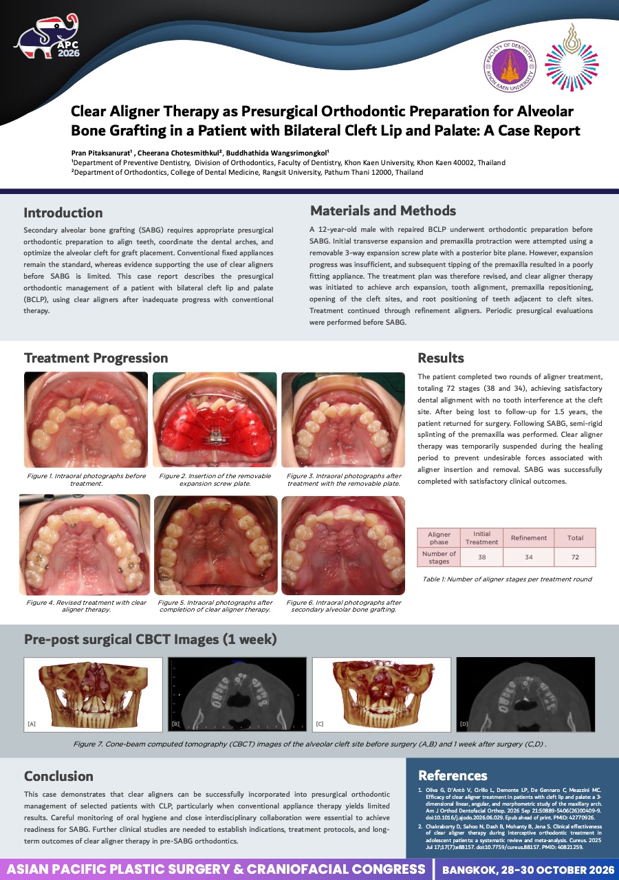
A 12-year-old male with repaired BCLP underwent presurgical orthodontic preparation before SABG. Initial transverse expansion and premaxillary protraction were attempted using a removable 3-way expansion screw plate with a posterior bite plane. Expansion was inadequate, and severe premaxillary tipping resulted in poor appliance fit. Rather than transitioning to fixed appliances, treatment was revised to clear aligners to achieve arch expansion, tooth alignment, premaxillary repositioning, cleft-site opening, and root uprighting adjacent to the clefts. Periodic presurgical evaluations were performed.
Results
The patient completed 2 rounds of aligner therapy comprising 72 stages (38 and 34), achieving satisfactory dental alignment and arch coordination for SABG. After a 1.5-year loss to follow-up, the patient returned for surgery. Following SABG, the premaxilla was semi-rigidly splinted, and aligner therapy was temporarily suspended during healing to prevent jiggling forces from aligner insertion and removal. SABG was completed successfully with satisfactory clinical outcomes.
Conclusion
This case demonstrates that clear aligners can be incorporated into presurgical orthodontic management of selected patients with BCLP when conventional appliance therapy yields limited results. Strict oral hygiene and interdisciplinary coordination were essential for graft readiness and postsurgical stability. Further studies are needed to establish indications, treatment protocols, and long-term outcomes of clear aligner therapy before SABG.
View only · Double-click poster to enlarge · Download & print disabled
Loading poster…
CP-0004
- Title
- Pre-Surgical Infant Orthopedic with Digital Nasal Stents and Sequential Alveolar Aligners in Unilateral Complete Cleft Lip and Palate: A Case Report
- Author(s)
- Pornpat Butpun
- Affiliation
- Division of Orthodontics, Faculty of Dentistry, Khon Kaen University, Khon Kaen, Thailand
Pre-Surgical Infant Orthopedic with Digital Nasal Stents and Sequential Alveolar Aligners in Unilateral Complete Cleft Lip and Palate: A Case Report CP-0004
Pornpat Butpun · Case Study
Introduction: Digital workflows and 3-dimensional (3D) printing have modernized presurgical infant orthopedics. The Digital Nasal Stents (DNS), developed and patented under the Faculty of Dentistry, Khon Kaen University, offers prefabricated, standardized nasal retainers for correcting cleft nose deformities. Complementing this, the AccuCleft system employs proprietary software to design sequential 3D-printed intraoral plates that accommodate maxillary growth, providing an efficient solution for large alveolar gaps (>12 mm) or patients with limited appointment availability.
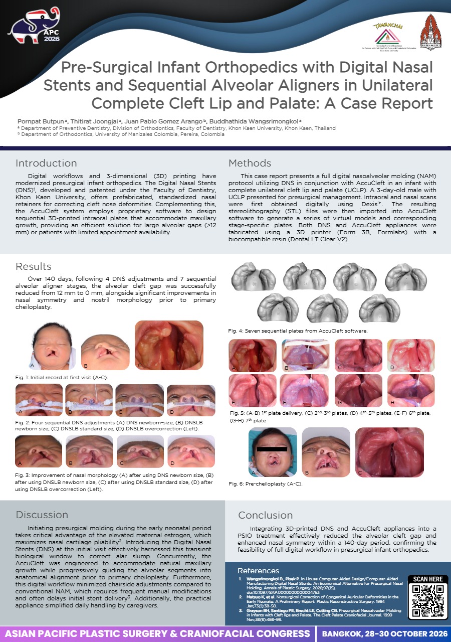
Methods: This case report presents a fully digital nasoalveolar molding (NAM) protocol utilizing DNS in conjunction with AccuCleft in an infant with complete unilateral cleft lip and palate (UCLP). A 3-day-old male with complete UCLP presented for presurgical management. Intraoral impressions and nasal scans were first obtained digitally using Dexis™. The resulting stereolithography (STL) files were then imported into AccuCleft software to generate a series of virtual models and corresponding stage-specific plates. Both DNS and AccuCleft appliances were fabricated using a 3D printer (Form 3B, Formlabs) with a biocompatible resin (Dental LT Clear V2).
Results: Over 140 days, following 4 DNS adjustments and 7 sequential alveolar aligner stages, the alveolar cleft gap was successfully reduced from 12 mm to 0 mm, alongside significant improvements in nasal symmetry and nostril morphology prior to primary cheiloplasty.
Conclusion: Integrating 3D-printed DNS and AccuCleft appliances into a digital NAM workflow effectively eliminated the alveolar cleft gap and enhanced nasal symmetry within a 140-day treatment window, confirming the feasibility of fully digital workflows in presurgical infant orthopedics management.
View only · Double-click poster to enlarge · Download & print disabled
Loading poster…
CP-0027
- Title
- From Cleft to Contour: Post-operative Nasal Molding Using The PSU Nasal Clip in a Patient with Cleft lip and Alveolus
- Author(s)
- PANUMAT INTHAMON
- Affiliation
- Center of Excellence in Correction and Rehabilitation of Dentofacial Deformities Faculty of Dentistry, Prince of Songkla University, Hat Yai, Thailand
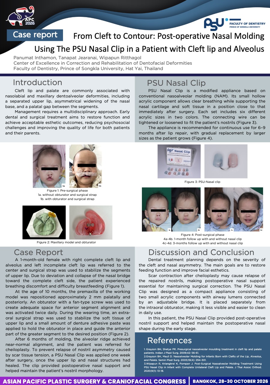
From Cleft to Contour: Post-operative Nasal Molding Using The PSU Nasal Clip in a Patient with Cleft lip and Alveolus CP-0027
PANUMAT INTHAMON · Case Study
Introduction
Cleft lip are associated with nasolabial and maxillary dentoalveolar defects. Early dental and surgical treatment aims to restore function and achieve acceptable esthetic outcomes, reducing psychosocial challenges and improving the quality of life for both patients and their parents.
Materials and methods: Case report
A 1-month-old female with right complete cleft lip and alveolus and left incomplete cleft lip was referred to the center. Due to collapse of the nasal bridge toward the complete cleft side, the patient experienced breathing discomfort and difficulty breastfeeding.
After 6 months of molding, the alveolar ridge achieved near-normal alignment, and the patient was referred for cheiloplasty.
To prevent relapse of nostril, a PSU Nasal Clip was applied one week after surgery, once the upper lip and nasal structures had healed. The clip provided postoperative nasal support and maintain the patient’s nostril morphology. The PSU Nasal Clip has small hollow acrylic component allows clear breathing while supporting the nasal cartilage and soft tissue in a position. Each set includes 6 different acrylic sizes in 2 colors. The connecting wire can be adjusted to fit the nostrils. The appliance is recommended for continuous use for 6–9 months after lip repair, with gradual replacement by larger sizes as the patient grows.
Conclusion
Dental treatment planning depends on the severity of the cleft and nasal asymmetry. The main goals are to restore feeding function and esthetics. Scar contraction after cheiloplasty may cause relapse, making postoperative nasal support essential for maintaining surgical correction.
The PSU Nasal Clip was designed as a compact appliance. It is placed separately from the intraoral obturator, making it less visible and easier to clean in daily use. The PSU Nasal Clip provided post-operative nostril support and maintain the postoperative nasal shape.
View only · Double-click poster to enlarge · Download & print disabled
Loading poster…
CP-0007
- Title
- Long-Term Outcomes of Korat Nasoalveolar Molding in a Patient with Complete Unilateral Cleft Lip and Palate: A 16-Years Interdisciplinary Follow-up
- Author(s)
- Somsri Klachoho
- Affiliation
- Cleft Center Maharat Nakhon Ratchasima Hospital
Long-Term Outcomes of Korat Nasoalveolar Molding in a Patient with Complete Unilateral Cleft Lip and Palate: A 16-Years Interdisciplinary Follow-up CP-0007
Somsri Klachoho · Case Study
Introduction:
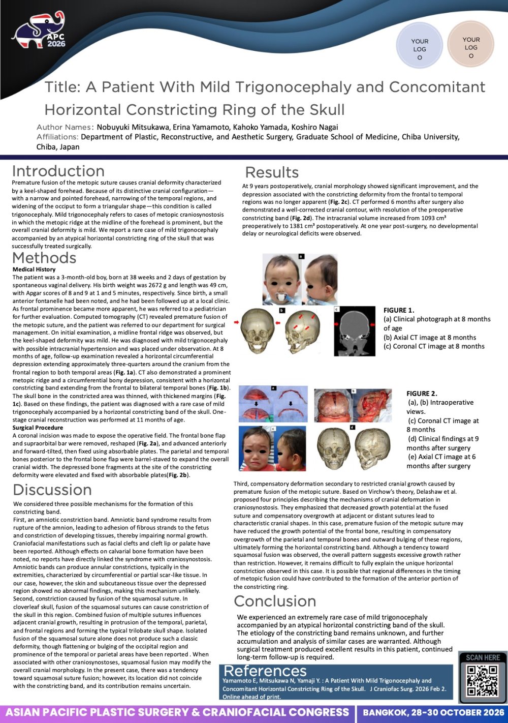
To present the comprehensive interdisciplinary treatment protocol and long-term outcomes of a patient with unilateral cleft lip and palate (UCLP) using the Korat Nasoalveolar Molding (Korat-NAM) protocol from infancy through adolescence.
Materials & Methods:
A 4-day-old male infant presented with a right complete UCLP, wide alveolar and palatal cleft and severe cleft lip nasal deformity. Korat-NAM-I was initiated immediately after birth to approximate the alveolar segments, reduce the alveolar and palatal clefts, elevate the collapsed nasal dome, improve columellar length, and mold the nasal cartilage. Primary cleft-lip repair with nasal-floor closure and primary rhinoplasty was performed at 6-months-old. Korat-NAM-II was worn until primary palatoplasty at 18 months -old to maintain nasal correction, followed by Korat-NAM-III from 18 months to further preserve nasal morphology. At 7.6-year-old, a fan-shaped maxillary-expansion-appliance was used to correct mid-arch collapse in preparation for secondary alveolar bone grafting. Comprehensive orthodontic treatment with extraction of the four first premolars using fixed appliances commenced at 13.4-year-old to resolve malocclusion. Speech therapy was provided continuously throughout growth.
Results:
At 15.8-year-old, patient demonstrated a straight facial profile with good nasal-projection, satisfactory lip and nasal esthetics, Class I occlusion, and 1-mm overjet. Serial lateral-cephalometric analysis from 7.6-year-old to 15.8-year-old showed ANB angle change from 2° to −1°, reflecting mandibula- growth while maintaining acceptable skeletal and dental relationships. Functional, esthetics and speech outcomes were satisfactory.
Discussion:
Korat-NAM represents a critical first step in the interdisciplinary management of complete UCLP. Early correction of alveolar and nasal deformities facilitates subsequent surgical, orthodontic, and speech interventions, reducing treatment complexity and the need for secondary surgical procedures.
Conclusions:
This case demonstrates that early nasoalveolar molding, integrated with coordinated interdisciplinary protocol, can achieve favorable functional and esthetic outcomes that are maintained through adolescence.
View only · Double-click poster to enlarge · Download & print disabled
Loading poster…
CP-0013
- Title
- Long-term Speech and Resonance Outcomes of Children with Unilateral Cleft Lip and Palate Born During the COVID-19 Pandemic: A Case Series from Northeast Thailand
- Author(s)
- Natthasart Unasri
- Affiliation
- Khon Kaen Hospital, Khon Kaen, Thailand
Long-term Speech and Resonance Outcomes of Children with Unilateral Cleft Lip and Palate Born During the COVID-19 Pandemic: A Case Series from Northeast Thailand CP-0013
Natthasart Unasri · Case Study
Background: COVID-19 disruptions delayed cleft surgeries and therapy. Long-term speech outcomes of pandemic-born cohorts remain underreported.
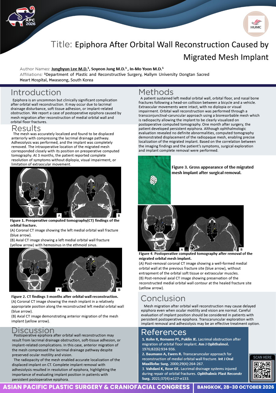
Objectives: To compare 2026 speech, resonance, and oral-motor outcomes of three pediatric patients with Left Unilateral Cleft Lip and Palate (UCLP) managed during 2020–2022 at Khon Kaen Hospital.
Methods: Retrospective tracking of three children (aged 5–6 years). Perceptual speech, resonance, and oral-motor function (Range of Motion; ROM) were evaluated alongside clinical timelines and complications.
Results: Outcomes in 2026 were highly heterogeneous:
Case 1 (High therapy adherence): Achieved competent sentence production, Mild articulation impairment, Fair intelligibility, and normal resonance (Hypernasality: 0, Nasal Emission: 0).
Case 2 (Late-onset hearing loss): Showed Moderate articulation impairment, Poor intelligibility, and mild resonance deficits (+1).
Case 3 (Surgical delay + oronasal fistula): Displayed restricted language (words/phrases), Severe articulation impairment, Poor intelligibility, resolved hypernasality (0), but persistent nasal emission (+1).
All cases showed "Adequate / Partial ROM".
Conclusion: Pandemic disruptions severely impacted long-term speech outcomes, heavily dictated by surgical timing, therapy compliance, fistulas, and hearing barriers. Targeted interventions are crucial to bridge these pandemic-induced care gaps.
View only · Double-click poster to enlarge · Download & print disabled
Loading poster…
Page 2 of 2 (21 poster(s))
Double-click or press Esc to close · Scroll to zoom